 Skip to content

Outer elbow pain can make simple daily activities—such as lifting a cup of tea, turning a doorknob, typing on a keyboard, or shaking hands—unbearably painful. Often referred to as Tennis Elbow, or medically as lateral epicondylitis, this condition is one of the most common reasons patients seek orthopedic care. Despite its athletic-sounding name, tennis elbow affects not just racket sports players, but anyone who performs repetitive wrist and forearm movements in their work or daily lives.
If you are looking for effective tennis elbow treatment in Varanasi, understanding the underlying causes, recognizing the symptoms, and exploring modern treatment approaches can help you make an informed decision about your care. Early orthopedic evaluation and structured rehabilitation are critical to preventing chronic tendon degeneration and regaining pain-free function.
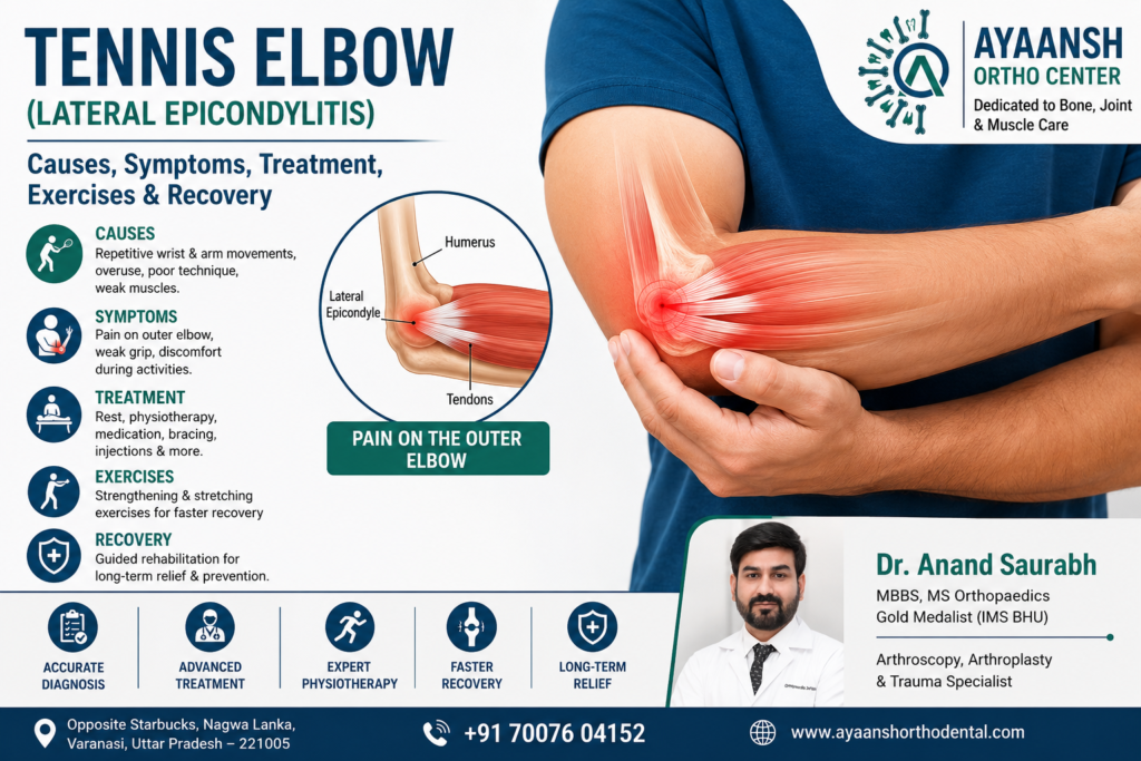
What is Tennis Elbow? Anatomy of the Elbow and Forearm
To understand tennis elbow, it is helpful to look at the anatomy of the forearm and elbow joint. The elbow is a hinge joint made up of three bones: the humerus (upper arm bone), the radius, and the ulna (forearm bones).
On the outer side of the humerus, there is a bony bump called the lateral epicondyle. This bony prominence serves as the attachment point for several muscles that extend the wrist and fingers. These muscles are connected to the bone via tough bands of tissue called tendons.
The primary muscle involved in tennis elbow is the Extensor Carpi Radialis Brevis (ECRB).
- The Role of the ECRB: The ECRB muscle helps stabilize the wrist when the elbow is straight. This is essential for activities like gripping or lifting.
- The Injury Mechanism: When the ECRB is overused, microscopic tears form in the tendon where it attaches to the lateral epicondyle. This triggers a cascade of inflammation and localized pain.
- Tendinitis vs. Tendinosis: While the condition is commonly referred to as “tendinitis” (implying active inflammation), chronic cases often exhibit “tendinosis.” Tendinosis is a degenerative state characterized by collagen breakdown, disorganized tissue fibers, and an abnormal increase in blood vessels (neovascularization), rather than active inflammatory cells. This is why long-standing tennis elbow fails to respond to simple anti-inflammatory medications and requires a more comprehensive approach to tissue healing.
Left untreated, these microscopic tears can coalesce into larger, painful partial-thickness or full-thickness tears of the common extensor tendon, leading to progressive muscle weakness and joint dysfunction.

Comprehensive Causes and Overuse Mechanisms
While tennis elbow is historically linked to racket sports, it is primarily a repetitive strain injury (RSI) that can be triggered by a wide range of athletic, occupational, and domestic activities.
1. Sports Kinematics and Training Errors
In sports, tennis elbow is usually caused by repetitive wrist extension against resistance. Common athletic causes include:
- Improper Equipment: Using a tennis racket that is too heavy, has an incorrect grip size, or has strings that are too tight.
- Poor Mechanics: A faulty backhand technique where the wrist bends excessively instead of remaining firm, placing sudden, high-velocity strain on the ECRB tendon.
- Sudden Intensity Spikes: Rapidly increasing playing time, training volume, or switching to heavier balls without letting the forearm muscles adapt.
2. Occupational Strain
People whose jobs require repetitive, forceful, or awkward forearm and wrist movements are at high risk. These occupations include:
- Plumbers, Carpenters, and Painters: Utilizing manual tools, painting walls, or turning wrenches requires repetitive gripping and twisting.
- IT Professionals and Office Workers: Prolonged typing and using a computer mouse without proper ergonomic support can overload the wrist extensors.
- Chefs and Butchers: Repetitive chopping, lifting heavy pans, and slicing meat put constant stress on the elbow tendons.
- Assembly Line and Factory Workers: Continuous manual sorting or assembly tasks involving repetitive wrist flexion and extension.
3. Domestic and Daily Activities
Simple household activities can also trigger or worsen lateral epicondylitis, such as:
- Extensive gardening or weeding.
- Wringing wet clothes or scrubbing floors.
- Using manual scissors or screwdrivers for home DIY projects.
- Carrying heavy grocery bags with an improper grip.
4. Systemic Risk Factors
Several internal factors can increase a person’s vulnerability to developing lateral epicondylitis:
- Age: It most commonly presents in adults between the ages of 30 and 50, as natural tendon elasticity and blood supply decrease with age.
- Smoking: Tobacco use impairs systemic microcirculation, slowing down the natural repair of microscopic tendon tears.
- Diabetes and Metabolic Syndrome: High blood sugar levels can alter the structural integrity of collagen, making tendons stiffer and more prone to micro-tearing.
- Poor Shoulder Mechanics: Weakness in the scapular stabilizers or a history of rotator cuff issues can cause a person to overcompensate by using their forearm muscles excessively. If you are experiencing shoulder issues alongside elbow pain, you can read our detailed guide on Rotator Cuff Tear Management to understand how upper-body biomechanics are linked.

Stages of Severity and Recognisable Symptoms
The symptoms of tennis elbow usually develop gradually. At first, you might notice a mild, occasional ache after a long day of work or physical activity. Over weeks or months, this can progress to a severe, constant pain that interferes with basic movements.
Stages of Lateral Epicondylitis
- Stage I (Mild): Mild pain or soreness after prolonged activity. The pain usually subsides with rest and basic home care.
- Stage II (Moderate): Pain occurs during the activity and persists for several hours afterward. It may begin to affect grip strength and performance.
- Stage III (Severe): Consistent pain during daily activities, even without heavy lifting. Pain may be present at rest and disrupt sleep.
- Stage IV (Chronic Degenerative): Continuous, severe pain accompanied by structural changes, marked weakness, and resistance to standard conservative therapies.
Characteristic Symptoms:
- Outer Elbow Tenderness: Intense tenderness when pressing directly on the lateral epicondyle (the bony bump on the outside of your elbow).
- Radiating Forearm Pain: A dull, aching pain that starts at the elbow and radiates down into the forearm and wrist.
- Weakness in Grip Strength: Notable difficulty when holding a glass, gripping a steering wheel, shaking hands, or holding a racket.
- Functional Limitation: Pain triggered by basic forearm rotation (pronation and supination), such as turning a key, opening a jar, or twisting a doorknob.
- Morning Stiffness: The elbow joint may feel stiff and sore upon waking, gradually loosening with gentle movement.
Differential Diagnosis: Ruling Out Other Elbow Conditions
Not all outer elbow pain is tennis elbow. It is important to get an accurate clinical evaluation from an experienced orthopedic specialist to rule out other potential diagnoses:
1. Golfer’s Elbow (Medial Epicondylitis)
While tennis elbow affects the outer side of the joint, golfer’s elbow affects the inner side, involving the wrist flexor tendons. The causes and treatment principles are similar, but the exact target muscles and exercises differ.
2. Radial Tunnel Syndrome
This condition is caused by the compression of the deep branch of the radial nerve (posterior interosseous nerve) as it passes through the forearm muscles near the elbow. The symptoms are very similar to tennis elbow, but the pain is typically localized about 2 inches further down the forearm and is characterized by a deep, aching muscle pain rather than bony tenderness.
3. Cervical Radiculopathy (Referred Neck Pain)
Compression of the C6 or C7 nerve roots in the neck can cause radiating pain, numbness, and tingling down the arm into the outer elbow and hand. This is often linked to underlying neck issues. If you suffer from chronic neck stiffness alongside elbow pain, explore our comprehensive resource on Cervical Spondylosis Treatment or check for signs of a Slip Disc.
4. Elbow Joint Osteoarthritis
Degeneration of the articular cartilage within the elbow joint itself can cause pain, clicking, popping, and a restricted range of motion. Unlike tennis elbow, joint arthritis pain is felt deep within the joint rather than specifically on the outer bony prominence.

Clinical Examination and Diagnostic Imaging
A precise diagnosis of tennis elbow begins with a physical examination by an orthopedic surgeon. During the clinical evaluation, several stress tests are performed to reproduce your symptoms and confirm the diagnosis:
- Cozen’s Test: The doctor stabilizes your elbow, asks you to make a fist, pronate your forearm, and extend your wrist against resistance. If this reproduces sharp pain at the lateral epicondyle, the test is positive.
- Mill’s Test: The doctor asks you to straighten your elbow fully while passively flexing your wrist and pronating your forearm. A positive test is indicated by pain at the outer elbow.
- Maudsley’s Test: The doctor applies resistance as you try to extend your middle finger. Pain during this movement indicates stress on the extensor digitorum and ECRB muscles.
Diagnostic Imaging
To confirm the diagnosis and assess the health of the joint and tendons, the following imaging modalities may be recommended:
- Digital X-rays: While X-rays do not show soft tissue like tendons, they are essential to rule out bone spurs, joint calcification, arthritis, or old fractures.
- Musculoskeletal Ultrasound: A dynamic, cost-effective imaging tool that allows the specialist to visualize the ECRB tendon in real-time. It can reveal thickening, focal hypoechogenicity (fluid), or microscopic tear patterns.
- MRI (Magnetic Resonance Imaging): If symptoms fail to improve after conservative treatment, an MRI provides high-contrast, multi-planar views. This helps evaluate the exact size and depth of any tendon tears, assess the articular cartilage, and rule out any intra-articular pathology.
Advanced Non-Surgical Treatment Strategies
Most patients with tennis elbow recover successfully without surgery. A structured, evidence-based approach is used to relieve pain, promote tendon healing, and restore full function.
1. The Acute Phase: Pain Relief and Protection
- Rest and Modification: This is the most crucial first step. Avoid activities that trigger pain. Modify daily tasks to reduce repetitive wrist extension.
- Cryotherapy (Ice Application): Apply ice packs wrapped in a thin towel for 15 minutes, 3 to 4 times a day, to manage acute discomfort and swelling.
- Medications: Short-term oral non-steroidal anti-inflammatory drugs (NSAIDs) or topical gels can help manage acute pain. However, they should not be relied upon for long-term recovery, as chronic tennis elbow is a degenerative rather than purely inflammatory condition.
2. Orthotic Support (Bracing and Splinting)
- Counterforce Brace: This is a specialized, adjustable strap worn about 2 inches below the elbow joint. It compresses the forearm muscles, absorbing and redistributing forces before they reach the damaged tendon attachment.
- Wrist Splint: In severe cases, wearing a wrist splint at night keeps the wrist in a neutral or slightly extended position. This rests the forearm extensor muscles and prevents involuntary wrist bending during sleep.
3. Regenerative Medicine: Platelet-Rich Plasma (PRP) Therapy
For patients with chronic, non-healing tennis elbow, Platelet-Rich Plasma (PRP) therapy has emerged as a highly effective, minimally invasive treatment option.
- How It Works: A small sample of the patient’s own blood is drawn and spun in a centrifuge to separate and concentrate the platelets, which are rich in growth factors.
- The Procedure: Under precise ultrasound guidance, the concentrated PRP is injected directly into the damaged, degenerated area of the ECRB tendon.
- The Healing Response: The growth factors stimulate cell proliferation, collagen synthesis, and neovascularization. This helps transition the chronic, degenerative “tendinosis” back into an active, healthy healing phase.
4. Shockwave Therapy (ESWT)
Extracorporeal Shockwave Therapy (ESWT) delivers high-energy acoustic waves to the painful outer elbow area. These shockwaves create therapeutic micro-trauma in the chronic tendon tissue, stimulating local blood flow, accelerating cellular repair, and breaking down calcifications within the tendon.
5. Therapeutic Injections
- Corticosteroid Injections: Can provide rapid, significant pain relief for 4 to 6 weeks, allowing patients to participate in physical therapy. However, repeat steroid injections are discouraged because they can weaken the collagen structure of the tendon, increasing the risk of tendon tears.
- Prolotherapy: Involves injecting an irritant solution (typically a concentrated dextrose mixture) to stimulate a localized, natural healing cascade in the chronically injured tendon.

Step-by-Step Physiotherapy and Exercises
Physical therapy is the cornerstone of long-term recovery for tennis elbow. Once the acute pain is under control, a progressive rehabilitation program is essential to stretch, strengthen, and remodel the damaged ECRB tendon.
Phase 1: Stretching and Flexibility (Pain-Free Range)
- Wrist Extensor Stretch: Extend your injured arm straight out in front of you with your palm facing down. Use your other hand to gently bend your wrist downward until you feel a stretch across the top of your forearm. Hold this position for 20–30 seconds. Relax and repeat 3 times. Perform this 2–3 times a day.
- Wrist Flexor Stretch: Extend your arm with your palm facing up. Use your other hand to gently pull your fingers downward toward the ground. Hold for 20–30 seconds, relax, and repeat 3 times.
Phase 2: Isometric Strengthening (Static Load)
- Wrist Extension Isometric Hold: Rest your forearm on a table with your palm facing down and your hand flat. Place your other hand on top of your fingers. Attempt to lift your wrist upward while resisting the movement with your top hand, so no actual joint movement occurs. Hold this static contraction for 10 seconds. Relax and repeat 5–10 times. This stimulates tendon healing without irritating the joint.
Phase 3: Eccentric Strengthening (The Gold Standard)
Eccentric training—where the muscle elongates while under tension—is highly effective for treating chronic tendon injuries.
- Eccentric Wrist Drops: Sit down and rest your forearm on a table, allowing your wrist to hang over the edge while holding a light dumbbell (0.5 to 1 kg). Use your healthy hand to lift the weight up (concentric phase). Then, remove your healthy hand and slowly lower the weight using only your injured arm over a slow count of 4 to 5 seconds (eccentric phase). Perform 3 sets of 10–15 repetitions daily.
- Towel Twist: Hold a rolled-up dry towel with both hands in front of you. Slowly twist the towel as if wringing out water, alternating directions. This helps rebuild functional rotational strength in the forearm muscles.
Phase 4: Grip and Rotational Strengthening
- Fist Squeezes: Grip a soft stress ball, a piece of therapy putty, or a sponge. Squeeze firmly for 5 seconds and release. Complete 15 repetitions.
- Pronation and Supination Resistance: Rest your forearm on a flat surface, holding a light hammer or a dumbbell by the bottom handle. Slowly rotate your forearm to turn the palm up (supination), then slowly rotate it to turn the palm down (pronation). Perform 10 controlled repetitions in each direction.
Workstation Ergonomics and Activities to Avoid
To prevent re-injury and protect your healing tendon, make these key changes to your daily routine:
Workstation Adjustments
- Neutral Wrist Position: Position your keyboard and mouse so your wrists remain straight and do not bend upward or downward while typing.
- Ergonomic Accessories: Use a vertical ergonomic mouse, which keeps your forearm in a neutral, semi-pronated “handshake” position, significantly reducing the strain on the lateral epicondyle.
- Take Regular Breaks: Follow the 45-15 rule: for every 45 minutes of typing or manual work, take a 5-minute break to gently stretch your wrists and forearms.
Lifting Modifications
- Palm-Up Lifting: When picking up objects (even light items like a laptop or grocery bag), always lift with your palm facing upward (supination). This transfers the lifting force to the larger, stronger biceps and wrist flexor muscles, sparing the injured extensor tendons on the outside of your elbow.
- Keep Objects Close: Avoid reaching out to pick up objects with an extended arm; keep your elbows bent and close to your body while lifting.
When is Surgery Necessary? Surgical Interventions
If your symptoms do not improve after 6 to 12 months of systematic non-surgical care, your orthopedic specialist may discuss surgical options.
1. Surgical Techniques
- Open ECRB Debridement: A small incision is made over the lateral epicondyle. The surgeon identifies the degenerated, scarred portion of the ECRB tendon, carefully removes (debrides) it, and repairs any tears. The healthy portion of the tendon is then anchored back to the bone.
- Arthroscopic Elbow Surgery: A minimally invasive technique performed through 2 or 3 tiny keyhole incisions using an arthroscope (a small camera). The surgeon visualizes the inside of the elbow joint, removes the damaged tendon tissue from underneath, and addresses any intra-articular issues. This technique typically causes less damage to surrounding healthy tissue and can lead to a faster recovery.
2. Post-Operative Rehabilitation Timeline
- Weeks 1–2: The arm is placed in a splint or sling to protect the repaired tendon while allowing early, gentle finger and wrist movements.
- Weeks 3–6: The splint is removed, and progressive passive and active-assisted range-of-motion exercises are started under physical therapy supervision.
- Weeks 6–12: Light strengthening exercises are introduced, focusing on isometric and gentle eccentric movements.
- Months 3–6: Advanced strengthening, functional training, and a gradual return to sports, heavy manual labor, and full physical activity are permitted.
Recovery Timelines: What to Expect
| Severity / Treatment Approach | Average Healing Period | Main Therapeutic Goal |
| Mild / Acute Cases | 4 to 8 weeks | Control inflammation, rest the ECRB tendon, and complete gentle stretching. |
| Moderate / Chronic Cases | 3 to 6 months | Structured eccentric exercises, wear a counterforce brace, and make ergonomic adjustments. |
| Regenerative PRP Therapy | 2 to 4 months | Cellular regeneration, progressive stretching, and eccentric remodeling. |
| Surgical Repair (Open/Arthroscopic) | 4 to 6 months | Gradual tendon-to-bone healing, physical therapy, and joint mobilization. |

Frequently Asked Questions (FAQs)
Can tennis elbow heal completely without surgery?
Yes. Over 90% of patients with tennis elbow make a full recovery using non-surgical methods such as rest, custom physical therapy, ergonomic adjustments, counterforce bracing, and regenerative therapies like PRP.
What is the fastest way to get relief from tennis elbow?
In the acute phase, the fastest way to relieve pain is through rest, regular ice application, wearing a counterforce brace to reduce tendon tension, and using short-term topical anti-inflammatory gels. However, long-term relief requires progressive strengthening.
Should I wear a tennis elbow brace all day?
No. You should wear a counterforce brace during activities that strain your forearm, such as lifting, typing, gardening, or sports. Avoid wearing it while resting, sleeping, or when your arm is relaxed, as constant compression can restrict local blood circulation.
Is walking good for tennis elbow?
Yes, walking is an excellent way to maintain cardiovascular health and systemic circulation, which supports overall healing. Walking does not put stress on the forearm extensor muscles, making it a safe exercise during your recovery.
How do I know if my elbow pain is tennis elbow or a pinched nerve?
Tennis elbow pain is typically localized to the outer bony bump of the elbow and is worsened by gripping or bending your wrist backward. A pinched nerve (such as radial tunnel syndrome or a nerve root compression in the neck) often causes a deep ache further down the forearm, or tingling, numbness, and burning sensations that travel down to the fingers.
Which doctor should I consult for tennis elbow in Varanasi?
You should consult an orthopedic doctor or a sports medicine specialist who is experienced in managing tendon disorders, musculoskeletal ultrasound diagnosis, and advanced treatments like physical therapy and PRP therapy.
Expert Tennis Elbow Treatment in Varanasi at Ayaansh Ortho Center
For patients experiencing persistent, debilitating elbow pain, Ayaansh Ortho Center provides diagnostic and therapeutic care. Located opposite Starbucks, Nagwa Lanka, Varanasi, our clinic offers comprehensive orthopedic management for sports injuries, tendon disorders, joint pain, arthritis, complex trauma, and spinal conditions.
The orthopedic team at Ayaansh Ortho Center is led by Dr. Anand Saurabh (MBBS, MS Orthopaedics), a Gold Medalist from IMS BHU with over 10 years of clinical experience. Dr. Anand Saurabh specializes in arthroscopy, joint reconstruction, advanced trauma care, and evidence-based regenerative therapies (such as PRP).
At our center, we develop highly personalized, patient-centric treatment plans designed to relieve your pain, restore natural joint mechanics, and improve your long-term quality of life. Whether you need an accurate diagnosis, a structured physical therapy program, or advanced regenerative care, we are here to support your recovery.
If you are dealing with persistent elbow discomfort or other issues like Knee Pain or chronic back pain, do not ignore the symptoms. Contact us today to schedule a consultation with an experienced orthopedic specialist in Varanasi.
