 Skip to content
An Anterior Cruciate Ligament (ACL) tear is one of the most severe and common knee injuries. This injury frequently affects athletes and active individuals who participate in high-impact sports. Specifically, a torn ACL causes immediate instability, severe pain, and rapid swelling. Consequently, patients often find it difficult to walk or bear weight on the injured leg. Therefore, an early and precise orthopedic evaluation is essential to prevent chronic joint instability and long-term cartilage damage.
If you are looking for advanced ACL reconstruction surgery in Varanasi, understanding your treatment options is crucial. At Ayaansh Ortho Center, we specialize in minimally invasive arthroscopic knee surgeries to help you regain full joint function. In this detailed guide, we will analyze the causes, symptoms, diagnostic tests, surgical options, and recovery protocols for an ACL tear.
What is the ACL? Understanding Knee Anatomy
First, we must examine the anatomical structure of the knee joint. The knee is a complex hinge joint that relies on four primary ligaments for stability. These ligaments connect the thighbone (femur) to the shinbone (tibia).
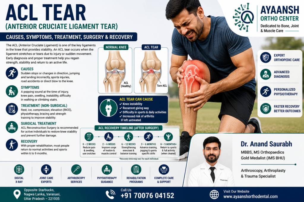
- Anterior Cruciate Ligament (ACL): This ligament runs diagonally in the middle of the knee. Specifically, it prevents the tibia from sliding out in front of the femur. It also provides critical rotational stability to the knee joint.
- Posterior Cruciate Ligament (PCL): This ligament runs parallel to the ACL. It prevents the tibia from sliding backward beneath the femur.
- Medial Collateral Ligament (MCL): Located on the inner side of the knee, this ligament prevents the joint from buckling inward.
- Lateral Collateral Ligament (LCL): Located on the outer side, this ligament prevents the joint from buckling outward.
Additionally, two wedge-shaped pieces of cartilage called menisci act as shock absorbers between the bones. Strong muscles, including the quadriceps and hamstrings, also support the joint.
During an ACL injury, the ligament undergoes excessive stretching or tearing. Because the ACL has a limited blood supply, it cannot heal on its own. Therefore, a complete tear typically requires surgical reconstruction to restore joint stability.

Common Causes of an ACL Tear
Generally, sudden, high-velocity forces cause an ACL tear. Surprisingly, about 70% of ACL injuries occur through non-contact mechanisms.
1. Non-Contact Biomechanical Forces
- Sudden Pivoting or Cutting: First, making a sharp, sudden change of direction while running is the most common cause. This movement places immense rotational stress on the knee.
- Awkward Landings: Second, landing poorly from a jump can force the knee into hyperextension or a knock-kneed position. Consequently, this sudden force tears the ligament fibers.
- Abrupt Deceleration: Third, stopping suddenly while running at high speeds can overload the ACL.
2. Direct Contact Trauma
- Collisions: A direct blow to the side of the knee can force the joint inward, tearing the ACL. For example, football tackles, rugby collisions, or motor vehicle accidents often cause this severe contact injury.
3. Sports Kinematics
Indeed, certain sports carry a much higher risk of ACL injuries. Specifically, football, basketball, soccer, skiing, and gymnastics require rapid deceleration and cutting. Consequently, athletes in these fields experience a high rate of ligament tears.

Recognizable Symptoms of an ACL Tear
When the ACL tears, the symptoms are usually dramatic and immediate. Therefore, most patients can pinpoint the exact moment the injury occurred.
- A Loud “Pop”: First, patients often hear or feel a loud popping sound inside the knee. This sound indicates the physical rupture of the ligament.
- Severe, Sudden Pain: Second, intense pain develops immediately. This pain makes it difficult or impossible to continue physical activity.
- Rapid Swelling (Hemarthrosis): Third, the knee swells significantly within 2 to 6 hours. This rapid swelling occurs because the ruptured ligament bleeds directly into the joint cavity.
- Feeling of Instability: Fourth, the knee feels unstable or “gives way” when you try to bear weight.
- Reduced Range of Motion: Finally, the accumulation of fluid makes it difficult to bend or straighten the knee fully.
If you ignore these symptoms, chronic instability can lead to secondary joint damage. Over time, an unstable knee can tear the meniscus cartilage. Consequently, this increases the risk of developing premature knee pain and osteoarthritis.
Associated Knee Injuries: The Unhappy Triad
Unfortunately, an ACL tear rarely occurs in isolation. High-force impacts often damage multiple structures within the knee joint simultaneously.
Specifically, orthopedic surgeons frequently identify a combination of injuries known as O’Donoghue’s Unhappy Triad. This complex injury includes:
- A complete ACL tear.
- A Medial Collateral Ligament (MCL) sprain or tear.
- A tear of the medial meniscus.
Consequently, this combination significantly increases joint instability. Therefore, the surgeon must address all damaged structures during treatment to ensure a successful recovery.

Clinical Examination and Diagnostic Tests
To confirm an ACL tear, an orthopedic surgeon must perform a detailed physical examination. During the exam, the doctor uses specific manual tests to check joint stability.
- Lachman Test: First, this is the most sensitive test for an ACL tear. The doctor bends your knee to 20–30 degrees. He then pulls the tibia forward while stabilizing the femur. Increased forward movement and a soft feel indicate a torn ligament.
- Anterior Drawer Test: Second, the doctor flexes your knee to 90 degrees. He then pulls the tibia forward. Excessive movement confirms ACL laxity.
- Pivot Shift Test: Third, this test evaluates the rotational stability of the joint. It is highly specific and helps assess the functional impact of the tear.
Diagnostic Imaging:
- Digital X-rays: First, X-rays help rule out bone fractures. They can also detect a Segond fracture. This is a specific bone avulsion on the outer side of the tibia that strongly indicates an ACL tear.
- MRI (Magnetic Resonance Imaging): Second, an MRI is the gold standard for soft tissue evaluation. Specifically, an MRI provides high-contrast images of the ACL, PCL, menisci, and collateral ligaments. Consequently, this scan allows the surgeon to plan the precise surgical approach.
Treatment Options: Non-Surgical vs. Surgical
Treatment for an ACL tear depends on the patient’s age, activity level, and the degree of knee instability.
1. Non-Surgical Treatment (Conservative Care)
Orthopedic specialists recommend conservative management only for specific patient groups. This includes older, less active individuals, or patients with minimal instability.
- Bracing: The patient wears a progressive knee brace to protect the joint from lateral movement.
- Physical Therapy: A structured program focuses on strengthening the quadriceps, hamstrings, and calf muscles. Consequently, these strong muscles help stabilize the joint.
- Activity Modification: The patient must avoid pivoting, cutting, or high-impact sports to prevent recurrent instability.
2. Surgical Treatment (ACL Reconstruction)
Active individuals, athletes, and patients with recurrent instability require surgical treatment. Because you cannot simply sew a torn ACL back together, the surgeon must reconstruct the ligament. Specifically, the surgeon replaces the torn ligament with a healthy tendon graft.

Detailed Look at ACL Reconstruction Surgery
Modern ACL surgery is highly advanced and minimally invasive. Specifically, surgeons perform keyhole arthroscopic surgery to reconstruct the ligament.
1. Graft Options (Autografts vs. Allografts)
The surgeon replaces the torn ACL using a tissue graft. There are three primary autograft options:
- Patellar Tendon (Bone-Patellar Tendon-Bone): The surgeon takes the middle third of the patellar tendon along with small bone plugs from the kneecap and shinbone. This graft offers strong bone-to-bone healing. Consequently, it is highly popular among professional athletes.
- Hamstring Tendon: The surgeon uses the semitendinosus and gracilis tendons from the back of the thigh. This graft requires smaller incisions and causes less pain in the front of the knee.
- Quadriceps Tendon: The surgeon takes a strip of the quadriceps tendon from above the kneecap. This graft provides a thick, strong alternative for revision surgeries or larger patients.
2. The Surgical Procedure
First, the surgeon makes 2 or 3 tiny keyhole incisions around the knee. He then inserts a small camera (arthroscope) to visualize the joint. Second, the surgeon removes the damaged ACL remnants and treats any associated meniscus tears. Third, the surgeon drills precise bone tunnels through the tibia and femur. Finally, he pulls the graft through these tunnels and secures it using specialized screws, buttons, or anchors.
Step-by-Step Rehabilitation and Recovery Timeline
Indeed, a successful outcome depends heavily on structured post-operative physical therapy. The rehabilitation process is criteria-based and progresses through distinct phases.
Phase 1: Protection and Joint Mobility (Weeks 1–4)
The primary goals are to reduce swelling, protect the healing graft, and regain full knee extension.
- Sling/Brace Support: The patient wears a locked knee brace and uses crutches to limit weight-bearing.
- Regaining Extension: It is critical to straighten the knee fully. The patient performs gentle heel props to allow gravity to stretch the joint.
- Quadriceps Activation: The patient performs straight leg raises and quad sets to prevent muscle wasting.
Phase 2: Progressive Weight-Bearing (Weeks 5–8)
During this phase, the patient transitions to full weight-bearing and begins building muscle strength.
- Weaning Off Crutches: The patient gradually discards crutches as gait mechanics normalize.
- Closed Kinetic Chain Exercises: The patient performs gentle wall slides, stationary cycling, and leg presses within a pain-free range of motion.
Phase 3: Muscle Strengthening and Balance (Weeks 9–16)
This phase focuses on building muscle volume and improving joint proprioception (balance).
- Squats and Lunges: The patient performs progressive squats and lunges to build quadriceps and gluteal strength.
- Proprioceptive Training: Using balance boards or foam pads helps train the neurological receptors within the new ligament.
Phase 4: Agility and Plyometrics (Months 4–6)
The patient prepares for dynamic movements.
- Straight-Line Jogging: Once muscle strength is adequate, the patient begins light, straight-line jogging.
- Shuttle Runs and Ladder Drills: These exercises introduce lateral movement and speed work.
Phase 5: Return to Sports (Months 9–12)
Before returning to competitive sports, the patient must pass rigorous functional tests. Specifically, the injured leg must demonstrate at least 90% of the strength and agility of the uninjured leg.

Long-Term Joint Health: Preventing Osteoarthritis
Undeniably, an ACL tear increases your risk of developing premature joint wear. Studies show that patients who suffer an ACL injury have a higher rate of knee osteoarthritis in later life.
However, you can minimize this risk by:
- Completing your physical therapy program fully.
- Maintaining a healthy body weight to reduce joint load.
- Continuing to perform knee-stabilizing exercises regularly.
If you experience ongoing joint wear, you can read our comprehensive resource on Knee Osteoarthritis Treatment to explore your long-term care options.
Expert ACL Reconstruction Surgery in Varanasi
If you need advanced ACL reconstruction surgery in Varanasi, choosing a specialized orthopedic center is critical. Ayaansh Ortho Center, located opposite Starbucks, Nagwa Lanka, Varanasi, offers comprehensive diagnostic and therapeutic services for sports injuries, ligament tears, and complex joint reconstructions.
Our orthopedic care is led by Dr. Anand Saurabh (MBBS, MS Orthopaedics), a Gold Medalist from IMS BHU with over 10 years of clinical experience. Dr. Anand Saurabh specializes in minimally invasive arthroscopy, sports medicine, joint replacement, and advanced trauma management. At our center, we combine surgical expertise with personalized rehabilitation programs to help you recover fully and safely.
