 Skip to content

The knee joint experiences significant daily stress. Specifically, it bears your entire body weight during walking, running, and jumping. To protect the bones from friction, the knee relies on specialized shock-absorbing cartilage structures called menisci. However, a sudden twist or gradual age-related wear can tear this protective cartilage. Consequently, a meniscus tear is one of the most common knee injuries. This injury causes localized pain, swelling, and mechanical joint restriction. Therefore, getting an early and accurate diagnosis is critical to preserving your knee joint over time.
If you are looking for advanced and evidence-based meniscus tear treatment in Varanasi, understanding your options is the first step toward recovery. At Ayaansh Ortho Center, our specialists provide personalized non-surgical and surgical care to restore joint mobility. In this comprehensive article, we will examine the anatomy, causes, symptoms, diagnostic tests, treatments, and rehabilitation phases of a meniscus tear.
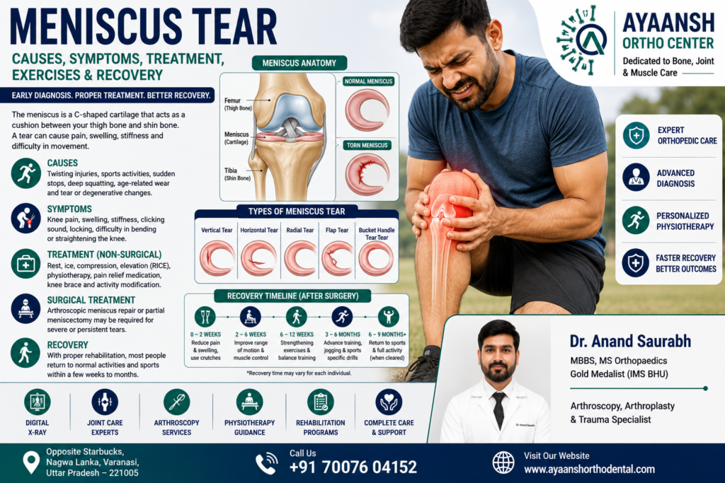
Understanding Knee Anatomy: What is the Meniscus?
First, we must examine the structure of the knee joint. The knee contains two C-shaped wedges of tough fibrocartilage located between the thighbone (femur) and the shinbone (tibia). These structures are the menisci.
1. Medial Meniscus
The medial meniscus sits on the inner side of the knee joint. Specifically, it is larger and C-shaped. Because it attaches firmly to the medial collateral ligament, it is relatively rigid. Consequently, the medial meniscus is more prone to tearing during injury.
2. Lateral Meniscus
The lateral meniscus sits on the outer side of the knee joint. It is smaller and more circular. Unlike the medial meniscus, it is highly mobile. Therefore, it is less susceptible to traumatic tears.
Biomechanical Functions of the Menisci:
- Shock Absorption: The menisci absorb physical shock during weight-bearing activities. Specifically, they distribute body weight across the entire joint.
- Joint Stability: They act as physical wedges to keep the rounded femur stable on the flat tibia.
- Lubrication and Nutrition: They help distribute synovial fluid across the joint. Consequently, this fluid lubricates the cartilage and provides vital nutrients.
The Vascular Zones: Red Zone vs. White Zone
To understand meniscus healing, we must look at the blood supply of the cartilage. The meniscus has a unique blood supply. Specifically, we divide the meniscus into three distinct zones.
1. The Red-Red Zone (Outer Third)
This outer region of the meniscus has a rich blood supply. Consequently, tears in this zone have excellent healing potential. Often, these outer tears can heal on their own. Alternatively, surgeons can successfully repair them using arthroscopic stitches.
2. The Red-White Zone (Middle Third)
This middle region has a marginal, limited blood supply. Healing in this zone is unpredictable. Therefore, the surgeon must evaluate each case carefully.
3. The White-White Zone (Inner Two-Thirds)
This inner region is completely avascular. Specifically, it has no blood supply. Consequently, tears in this zone cannot heal on their own. Similarly, surgical repair in this zone usually fails. Therefore, surgeons typically perform a partial meniscectomy to trim away the damaged tissue in this region.

Common Causes of a Meniscus Tear
Generally, meniscus tears happen through two primary mechanisms: acute trauma or gradual degeneration.
1. Traumatic Tears (Acute Injury)
First, acute tears usually occur during high-impact sports. Specifically, a sudden, forceful twist of the knee while the foot is planted can tear the fibrocartilage. Similarly, deep squatting or a direct blow to the knee can cause traumatic damage. Athletes in sports like football, basketball, tennis, and soccer are highly vulnerable to these injuries.
2. Degenerative Tears (Chronic Wear)
Second, older adults often develop degenerative tears. Over time, the meniscus tissue naturally loses its water content and elasticity. Consequently, the cartilage becomes thin, brittle, and weak. In these cases, simple daily activities can cause a tear. For example, rising from a low chair or twisting slightly while stepping out of a car can damage a degenerated meniscus.
Types of Meniscus Tears (Tear Morphology)
Indeed, meniscus tears can present in various physical shapes and patterns. Orthopedic surgeons classify these tears based on their appearance on diagnostic scans.
- Radial Tears: First, radial tears start at the inner, avascular margin and extend outward. These tears cannot heal on their own.
- Horizontal Tears: Second, horizontal tears run parallel to the fibers of the meniscus. They are common in older adults and often respond well to non-surgical care.
- Longitudinal Tears: Third, longitudinal tears run along the length of the meniscus. They often occur in the outer, vascular red-red zone. Consequently, they are excellent candidates for surgical repair.
- Bucket-Handle Tears: Fourth, this is a severe form of a longitudinal tear. Specifically, the torn portion of the cartilage flips inside the joint. This displacement can physically lock the knee, making it impossible to straighten.
- Flap Tears: Finally, a flap tear involves a small piece of cartilage peeling back. This loose flap can catch in the joint, causing painful clicking.

Recognizable Symptoms of a Meniscus Tear
When a meniscus tears, the symptoms vary based on the severity and type of the injury. Generally, the symptoms develop over several hours or days.
- Delayed Swelling and Stiffness: First, unlike an ACL tear which swells instantly, a meniscus tear causes gradual swelling. Typically, swelling develops over 24 to 36 hours.
- Localized Joint Line Pain: Second, patients experience pain directly along the joint line. Specifically, pressing on the inner or outer side of the knee will reproduce the pain.
- Mechanical Catching or Locking: Third, the knee may catch, pop, or physically lock during movement. This happens when the torn piece of cartilage interferes with normal joint motion.
- Inability to Bend or Straighten Fully: Fourth, the patient struggles to complete a full range of motion. Deep squatting is highly painful.
- Feeling of Instability: Finally, the knee may feel weak or “give way” during walking. This occurs as a protective muscle reflex to avoid pain.
Clinical Evaluation and Orthopedic Tests
To formulate a targeted treatment plan, an orthopedic surgeon must perform a detailed clinical evaluation. During the exam, the doctor will use physical stress tests to isolate the meniscus.
- McMurray’s Test: First, the doctor flexes your knee fully. He then rotates your tibia inward or outward while extending the joint. If a painful click or catch occurs along the joint line, the test is positive.
- Thessaly Test: Second, the patient stands on the injured leg with the knee bent to 20 degrees. The patient then rotates their body side-to-side. Increased pain or catching confirms a tear.
- Apley’s Compression Test: Third, the patient lies face down with the knee bent to 90 degrees. The doctor applies downward pressure while rotating the foot. Increased pain indicates meniscus damage.
Diagnostic Imaging:
- Digital X-rays: While X-rays cannot show soft cartilage, they are necessary to rule out arthritis, bone spurs, or fractures.
- MRI (Magnetic Resonance Imaging): An MRI is the gold standard for meniscus diagnosis. Specifically, it provides highly detailed, multi-planar images of the joint. Consequently, the scan reveals the exact size, location, and pattern of the tear.
Non-Surgical Treatment Options
Fortunately, many patients with small, stable meniscus tears do not require surgery. This is particularly true for tears located in the vascular outer third or degenerative tears.
1. The R.I.C.E. Protocol
- Rest: Avoid activities that require twisting, pivoting, or deep squatting.
- Ice: Apply an ice pack wrapped in a towel for 15 minutes, 3 to 4 times a day, to reduce acute swelling.
- Compression: Wear an elastic compression bandage to support the joint.
- Elevation: Prop your leg up above your heart level to minimize fluid accumulation.
2. Medications
Additionally, short-term oral anti-inflammatory drugs (NSAIDs) can help manage localized pain and swelling. However, these medications do not heal the cartilage. Therefore, you must combine them with physical therapy.
3. Progressive Physical Therapy
Furthermore, a customized physical therapy program is crucial. The therapist focuses on strengthening the quadriceps, hamstrings, and hip muscles. Consequently, these strong muscles absorb physical forces, reducing the load on the healing meniscus.
4. Regenerative Medicine (PRP Therapy)
For chronic, slow-healing tears in the red-white zone, Platelet-Rich Plasma (PRP) therapy is an advanced option. The doctor extracts concentrated growth factors from your own blood. He then injects them directly into the torn meniscus under ultrasound guidance. Consequently, this treatment stimulates cellular repair and collagen synthesis.

Surgical Treatment: Arthroscopic Meniscus Surgery
If non-surgical treatment fails to provide relief, or if the knee is mechanically locked, surgery is necessary. Today, surgeons perform these procedures using minimally invasive keyhole knee arthroscopy.
1. Arthroscopic Meniscal Repair
When possible, the surgeon prefers to repair and preserve the meniscus. This is highly common for tears in the outer, vascular red-red zone of young patients. Specifically, the surgeon uses tiny instruments and specialized sutures to stitch the torn cartilage back together. Consequently, this procedure preserves the joint’s natural shock-absorbing capacity. Therefore, it protects the joint from long-term wear and tear.
2. Arthroscopic Partial Meniscectomy
If the tear occurs in the avascular white-white zone, the tissue cannot heal. In this case, the surgeon performs a partial meniscectomy. Specifically, the surgeon trims away only the damaged, loose edges of the cartilage. He leaves behind as much healthy tissue as possible to maintain joint function. As a result, the procedure relieves pain and prevents catching or locking.
Rehabilitation Protocol and Recovery Timeline
Indeed, your recovery timeline depends heavily on the type of surgery performed.
Recovery After Partial Meniscectomy:
Because the surgeon removes the torn tissue, recovery is relatively quick.
- Weight-Bearing: The patient can bear weight as tolerated immediately after surgery. Crutches are usually necessary for only 3 to 5 days.
- Range of Motion: Gentle bending and straightening exercises start immediately.
- Return to Normal Activity: Most patients return to light daily tasks within 2 weeks. Full return to sports or manual work typically takes 4 to 6 weeks.
Recovery After Meniscal Repair:
Stitched cartilage requires strict protection to heal. Therefore, the recovery protocol is much more conservative.
- Immobilization: The patient wears a locked knee brace for 4 to 6 weeks.
- Restricted Weight-Bearing: The patient must use crutches. They must avoid putting full weight on the leg for the first 4 weeks.
- Controlled Flexion: The brace restricts knee bending to 90 degrees during the early healing phase to prevent stress on the stitches.
- Return to Normal Activity: Returning to daily tasks takes about 6 to 8 weeks. Full return to competitive sports or heavy lifting typically requires 4 to 6 months of systematic physical therapy.

Step-by-Step Rehabilitation Exercises
A structured physical therapy program is essential for a complete recovery. Exercises must progress in a controlled, pain-free manner.
Phase 1: Early Mobility and Quad Activation (Weeks 1–2)
- Quadriceps Sets: Sit with your leg straight out in front of you. Tighten your thigh muscle by pushing the back of your knee down into the floor. Hold for 5 seconds and relax. Repeat 15 times for 3 sets.
- Straight Leg Raises: Tighten your thigh muscle and slowly lift your leg 12 inches off the ground, keeping your knee straight. Hold for 3 seconds and slowly lower it. Perform 10 repetitions.
- Ankle Pumps: Move your foot up and down rapidly. This simple movement promotes lower-limb circulation and prevents blood clots.
Phase 2: Range of Motion and Light Strengthening (Weeks 3–6)
- Heel Slides: Sit on a flat surface. Slowly slide your heel toward your buttocks, bending your knee as far as comfortable. Slide it back out. Complete 15 repetitions.
- Stationary Cycling: Use a stationary bike with low resistance. This exercise improves joint fluid circulation and range of motion.
- Calf Raises: Stand tall, holding onto a chair for balance. Slowly lift up onto your toes, hold for 2 seconds, and lower back down. Perform 15 repetitions.
Phase 3: Advanced Strengthening and Balance (Weeks 7–12)
- Wall Squats: Lean your back flat against a wall. Step your feet 12 inches forward. Slowly slide down the wall until your knees are bent to 45 degrees. Hold for 5 seconds and slide back up. Complete 10 repetitions.
- Single-Leg Balance: Stand on your injured leg for 30 seconds to train the neurological balance receptors within the joint.
Prevention and Ergonomic Tips for Knee Safety
To protect your knee joint and prevent future meniscus tears, implement these preventive habits:
- Maintain a Healthy Body Weight: Extra body weight places significant stress on the knees. Losing weight reduces the cumulative load on the menisci.
- Strengthen the Leg Muscles: Perform regular exercises to strengthen your quadriceps, hamstrings, and gluteal muscles. Strong muscles stabilize the knee and absorb physical impacts.
- Avoid Sudden, Unprepared Pivoting: Warm up thoroughly before playing sports. Avoid making sudden changes of direction with your feet planted firmly on the ground.
- Use Proper Ergonomics: Avoid remaining in a deep squatting position for long periods. If your job requires squatting, take regular standing breaks to stretch your knees.

Expert Meniscus Tear Care at Ayaansh Ortho Center in Varanasi
If knee pain, clicking, or locking is affecting your daily life, seeking specialized care is critical. Ayaansh Ortho Center, located opposite Starbucks, Nagwa Lanka, Varanasi, provides advanced diagnostic and treatment services for knee disorders, ligament injuries, and complex cartilage tears.
Our orthopedic services are led by Dr. Anand Saurabh (MBBS, MS Orthopaedics), a Gold Medalist from IMS BHU with over 10 years of clinical experience. Dr. Anand Saurabh specializes in minimally invasive arthroscopy, joint reconstruction, sports medicine, and advanced trauma care. At our center, we develop personalized, patient-centric treatment plans to help you relieve pain, restore joint mobility, and return to your active lifestyle safely.
Do not let chronic knee pain limit your physical potential. If you require an accurate diagnosis or an expert orthopedic consultation, please contact our team to schedule an appointment.
