 Skip to content
The hip is one of the largest and most resilient weight-bearing joints in the human body. Specifically, it is a ball-and-socket joint that supports your upper body weight while allowing you to walk, run, climb, and bend. However, because the hip experiences constant mechanical stress, it is highly prone to wear, tear, and injury. Consequently, hip pain is a widespread issue that affects individuals of all age groups. It can severely restrict your mobility, disrupt your sleep, and lower your overall quality of life.
If you are looking for effective hip pain treatment in Varanasi, getting an early and accurate diagnosis is critical. At Ayaansh Ortho Center, our specialists provide comprehensive, personalized care to help you regain pain-free mobility. In this detailed article, we will examine the anatomy, causes, symptoms, diagnostic tests, treatments, and rehabilitation exercises for hip pain.
Anatomy of the Hip Joint
First, we must understand the anatomical structure of the hip. The hip is a ball-and-socket joint that connects the lower limbs to the pelvis.
- The Ball (Femoral Head): This is the rounded top of your thighbone (femur).
- The Socket (Acetabulum): This is the cup-like cavity in your pelvis bone where the femoral head fits.
- The Labrum: This is a ring of tough, flexible cartilage that lines the outer rim of the socket. Specifically, the labrum acts as a rubber seal to hold the ball securely inside the socket.
- Articular Cartilage: A smooth, slippery tissue covers both the femoral head and the acetabulum. This cartilage absorbs shock and prevents the bones from rubbing directly against each other.
- Synovial Membrane: This thin membrane surrounds the joint and produces synovial fluid. This fluid lubricates the joint, allowing smooth, friction-free movement.
- Muscles and Ligaments: Strong ligaments encase the joint to keep it stable. Additionally, major muscle groups—including the gluteals, hip flexors, adductors, and hamstrings—power the movements of the leg.

Classifying Hip Pain by Location
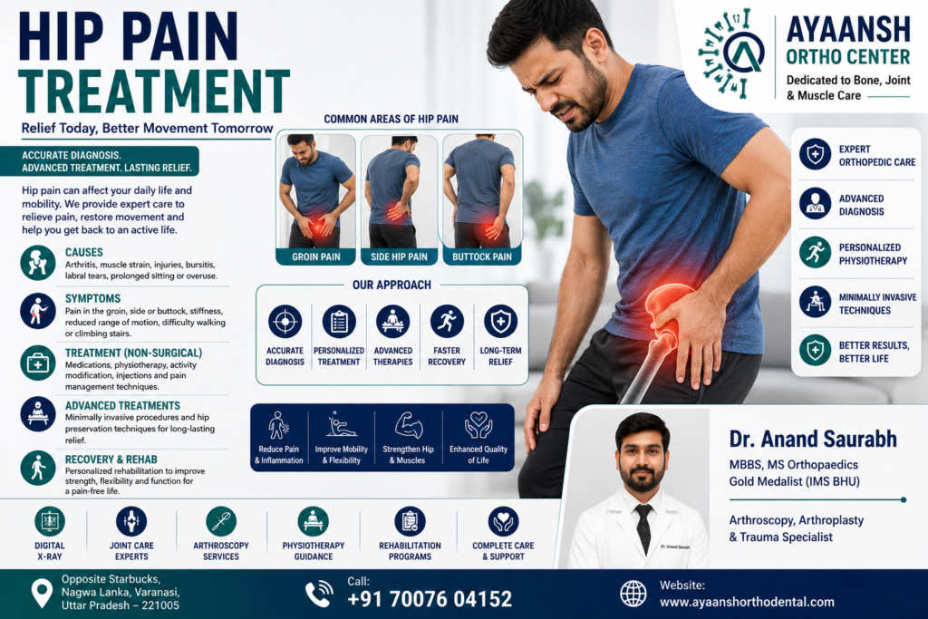
Indeed, the exact location of your hip pain provides vital clues regarding its underlying cause. Orthopedic specialists typically categorize hip pain into three primary regions.
1. Anterior Hip Pain (Front of the Hip or Groin)
Typically, pain felt on the inside of the hip or in the groin indicates a problem within the hip joint itself.
- Osteoarthritis: The gradual wear of the protective articular cartilage.
- Femoroacetabular Impingement (FAI): Structural abnormalities of the bones that cause friction in the joint.
- Hip Labral Tears: Damage or tears in the ring of cartilage lining the socket.
- Iliopsoas Tendinitis: Inflammation of the main hip flexor tendon in the front of the joint.
2. Lateral Hip Pain (Outer Side of the Hip)
Generally, pain on the outer side of the hip stems from issues in the soft tissues, muscles, tendons, or bursa surrounding the joint.
- Trochanteric Bursitis: Inflammation of the fluid-filled sac (bursa) over the outer bony point of the femur.
- Gluteal Tendinopathy: Irritation or micro-tearing of the gluteus medius or minimus tendons.
- Iliotibial (IT) Band Syndrome: Friction of the tight fibrous band running down the outer thigh.
3. Posterior Hip Pain (Buttock or Back of the Hip)
Often, posterior hip pain is linked to issues in the lower back, sacroiliac joints, or deep gluteal muscles.
- Sciatica and Lumbar Radiculopathy: Compression of the sciatic nerve in the lower back that radiates pain to the buttock.
- Sacroiliac (SI) Joint Dysfunction: Inflammation or abnormal movement in the joint connecting the pelvis to the spine.
- Piriformis Syndrome: Spasm of the deep piriformis muscle that compresses the sciatic nerve.

Primary Causes of Hip Pain
Generally, several acute injuries and chronic conditions can trigger hip pain. Understanding these causes helps guide the treatment plan.
1. Hip Osteoarthritis (Wear-and-Tear Arthritis)
First, osteoarthritis is the most common cause of chronic hip pain in older adults. Over time, the smooth articular cartilage wears away. Consequently, the bones begin to rub directly against each other. This friction causes bone spurs (osteophytes), localized inflammation, joint stiffness, and deep groin pain.
2. Avascular Necrosis (AVN) of the Hip
Second, Avascular Necrosis (AVN) is a serious condition that occurs when the blood supply to the femoral head is temporarily or permanently cut off. Without a healthy blood supply, the bone cells in the femoral head die. Consequently, the bone structure weakens, eventually causing the ball to collapse. AVN is common in young adults and is often linked to steroid use, excessive alcohol consumption, or trauma.
3. Hip Labral Tears
Third, trauma, repetitive twisting, or FAI can tear the labrum cartilage. When the labrum tears, it can flap inside the joint. As a result, patients feel a sharp pain, clicking, or catching sensation in the groin.
4. Trochanteric Bursitis (Greater Trochanteric Pain Syndrome)
Fourth, the greater trochanter is the bony bump on the outside of your thigh. A fluid-filled sac, called the bursa, sits over this bone to reduce friction. Repetitive strain or direct falls can inflame this bursa. Consequently, the patient experiences sharp pain on the outside of the hip, especially when lying on that side.
5. Femoroacetabular Impingement (FAI)
Fifth, FAI involves abnormal bone growth on either the femoral head (Cam impingement) or the acetabular rim (Pincer impingement). During movement, these abnormal bone structures pinch against each other. Consequently, this friction damages the labrum and joint cartilage over time.
6. Fractures and Trauma
Finally, hip fractures are major orthopedic emergencies, particularly in elderly individuals with osteoporosis. A sudden fall can break the femoral neck, requiring immediate surgical stabilization.
Common Symptoms of Hip Pathology
When the hip joint is damaged, the symptoms can vary from a dull ache to sharp, stabbing pain.
- Groin Pain: Pain located in the front groin crease is the hallmark symptom of an internal hip joint issue.
- Limping (Antalgic Gait): The patient alters their walking pattern to minimize weight-bearing on the painful leg.
- Joint Stiffness: Difficulty bending the hip, putting on socks, or getting out of a low car, especially in the morning.
- Reduced Range of Motion: Difficulty rotating the leg inward or outward.
- Clicking, Snapping, or Popping: A mechanical sensation or sound inside the joint during movement.
- Pain with Weight-Bearing: Pain that worsens during walking, climbing stairs, or standing for long periods.
- Referred Pain: Hip joint issues can sometimes refer pain down to the thigh or the knee joint.

Clinical Examination and Orthopedic Tests
To identify the exact source of your pain, an orthopedic specialist will perform several specific physical tests during the clinical evaluation.
- FABER Test (Patrick’s Test): First, the doctor asks you to lie on your back. He bends your knee, rotates your leg outward, and rests your ankle on your opposite thigh (forming a figure-4 position). He then applies gentle downward pressure. Groin pain indicates a hip joint issue, while back pain suggests an SI joint problem.
- FADIR Test: Second, the doctor flexes your hip, adducts the leg toward the midline, and rotates it inward. Pain in the groin crease strongly suggests FAI or a labral tear.
- Trendelenburg Test: Third, you stand on one leg. If your pelvis drops on the opposite side, it indicates weakness in the gluteal muscles of the standing leg.
- Log Roll Test: Finally, you lie flat on your back. The doctor gently rolls your entire leg inward and outward. Pain during this passive rotation suggests an active hip joint issue.
Diagnostic Imaging:
- Digital X-rays: First, X-rays are the primary tool used to check for joint space narrowing, bone spurs, FAI structures, and signs of AVN or fractures.
- MRI (Magnetic Resonance Imaging): Second, an MRI is essential for visualizing soft tissues. Specifically, it reveals labral tears, muscle strains, early-stage AVN, and bone marrow edema.
- Diagnostic Ultrasound: Finally, ultrasound helps detect trochanteric bursitis and fluid accumulation within the joint capsule.
Non-Surgical Treatment Options
Fortunately, many patients with hip pain can achieve complete relief through non-surgical, conservative treatments.
1. Activity Modification and Rest
Initially, you must rest the joint. Avoid activities that involve repetitive impact, such as running or jumping. Similarly, minimize climbing stairs or sitting in deep, low chairs to prevent joint compression.
2. Medications
Additionally, short-term use of oral anti-inflammatory drugs (NSAIDs) helps manage acute pain and localized inflammation. However, these medications do not address the structural cause of your pain. Therefore, you must combine them with other therapies.
3. Targeted Physical Therapy
Furthermore, a customized physical therapy program is the cornerstone of conservative care. The therapist focuses on strengthening the gluteal muscles, hamstrings, and core muscles. Consequently, these strong muscles support the pelvis, stabilize the hip joint, and reduce wear on the cartilage.
4. Intra-Articular Injections
If severe pain limits your mobility or physical therapy progress, the doctor may recommend an injection:
- Corticosteroid Injections: Under precise ultrasound or fluoroscopic guidance, the doctor injects a steroid directly into the hip joint or bursa. This provides powerful, localized pain and inflammation relief.
- PRP (Platelet-Rich Plasma) Therapy: This advanced regenerative option uses growth factors from your own blood. Under ultrasound guidance, the doctor injects PRP into damaged tendons or early arthritic joints to promote cellular healing.

Surgical Treatment: When is it Needed?
If conservative treatments fail to provide relief after several months, or if you have a severe structural issue, surgery may be necessary.
1. Hip Arthroscopy (Keyhole Surgery)
First, hip arthroscopy is a minimally invasive surgical option. The surgeon uses tiny incisions and a camera to access the joint. He can then trim or repair a torn labrum. He can also shave down abnormal bone growths (FAI) to restore smooth joint movement. This procedure is highly effective for active, younger patients.
2. Core Decompression
Second, for early-stage Avascular Necrosis (AVN), core decompression is a valuable joint-preserving option. The surgeon drills small holes into the dead area of the femoral head. This relieves pressure inside the bone and allows new blood vessels to grow. Consequently, it can halt the progression of AVN.
3. Total Hip Replacement (Total Hip Arthroplasty)
Finally, for patients with severe, end-stage osteoarthritis or collapsed AVN, a total hip replacement is highly successful.
- The Procedure: The surgeon removes the damaged femoral head and socket lining. He then replaces them with biocompatible artificial implants (typically ceramic, metal, and highly durable polyethylene).
- The Outcome: This surgery provides dramatic pain relief and restores excellent, near-natural joint mobility, allowing patients to return to daily activities comfortably.
Step-by-Step Rehabilitation Exercises
A structured, low-impact exercise program is crucial for maintaining hip joint mobility and strength. Please consult an orthopedic surgeon or physical therapist before starting these exercises.
Phase 1: Gentle Flexibility and Mobility
- Gluteal Sets: Lie on your back with your knees bent. Squeeze your buttock muscles tightly, holding for 5 seconds. Repeat 15 times for 3 sets.
- Hip Flexor Stretch: Kneel on your injured knee, with your opposite foot flat on the floor in front of you. Gently lean your hips forward until you feel a stretch in the front of your hip. Hold for 20 seconds. Repeat 3 times.
Phase 2: Isometric and Core Stability
- Clamshells: Lie on your side with your hips and knees bent at 45 degrees. Keep your feet touching. Slowly lift your top knee as high as possible without moving your pelvis. Hold for 2 seconds and lower. Complete 15 repetitions. This targets the stabilizing gluteus medius muscle.
- Hip Bridges: Lie on your back with your knees bent and feet flat on the floor. Slowly lift your hips toward the ceiling until your body forms a straight line from your shoulders to your knees. Hold for 3 seconds and lower. Repeat 12 times.
Phase 3: Advanced Functional Strengthening
- Standing Hip Abduction: Stand tall, holding onto a chair for balance. Slowly lift your injured leg outward to the side, keeping your knee straight. Hold for 2 seconds and return. Complete 15 repetitions.
- Bird-Dog Exercise: Kneel on your hands and knees. Slowly extend your right arm forward and your left leg backward. Hold for 3 seconds. Return and repeat on the opposite side. This strengthens your core and lower back, protecting the pelvis.
Preventive Measures for Long-Term Hip Health
To protect your hip joints from premature wear, implement these proactive daily habits:
- Maintain a Healthy Weight: Every extra pound adds significant load to your hips. Maintaining a healthy weight directly reduces joint stress.
- Avoid Repetitive Impact: If you have early hip discomfort, transition from high-impact running to low-impact swimming, cycling, or elliptical training.
- Use Proper Ergonomics: Sit in chairs that keep your hips higher than your knees. This prevents joint pinching and groin compression.
- Stretch Regularly: Keep your hip flexors, hamstrings, and gluteal muscles flexible to ensure balanced joint movement.

Expert Hip Care at Ayaansh Ortho Center in Varanasi
If persistent hip pain is limiting your mobility or daily tasks, seeking specialized care is essential. Ayaansh Ortho Center, located opposite Starbucks, Nagwa Lanka, Varanasi, offers comprehensive diagnostic and treatment services for hip disorders, joint replacement, sports injuries, and spinal conditions.
Our orthopedic care is led by Dr. Anand Saurabh (MBBS, MS Orthopaedics), a Gold Medalist from IMS BHU with over 10 years of clinical experience. Dr. Anand Saurabh specializes in hip joint replacement, arthroscopy, trauma care, and evidence-based regenerative medicine. At our center, we focus on providing personalized, patient-centric treatment plans to help you relieve pain, restore joint biomechanics, and safely return to your active lifestyle.
Do not let chronic hip pain restrict your active life. If you require an accurate diagnosis, a second opinion, or a professional orthopedic consultation, contact our clinic today to book an appointment.
External References
- American Academy of Orthopaedic Surgeons (AAOS) – Hip Pain Guide
- Mayo Clinic – Hip Pain Symptoms & Causes
- Cleveland Clinic – Hip Pain Causes & Diagnostic Overview
