 Skip to content

Persistent joint stiffness, localized swelling, or a sharp ache in your kneecap can severely limit your physical freedom. Because the knee joint bears your entire body weight during movement, it is highly prone to injuries and wear. Whether your discomfort stems from a sudden sports injury or gradual age-related cartilage degeneration, seeking professional care is essential. Consequently, if you are looking to eliminate joint discomfort and restore your active life, seeking the right Knee Pain Treatment in Varanasi can help you achieve a safe and successful recovery.
At Ayaansh Ortho Center, our metabolic bone and joint care team specializes in comprehensive diagnostic screenings and advanced knee therapies. We combine digital diagnostic scanning, personalized physical therapy, and evidence-based medical treatments to manage joint pain. In this highly comprehensive, clinically detailed guide, we will analyze the complex anatomy of the knee, its primary causes of pain, recognizable symptoms, advanced diagnostic tests, non-surgical and surgical options, and preventative exercises.
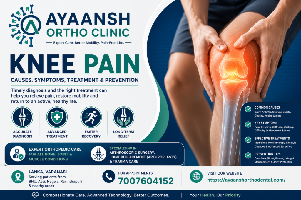
Finding the Best Knee Pain Treatment in Varanasi
Every knee joint undergoes unique mechanical and structural changes during wear or trauma. Therefore, choosing specialized Knee Pain Treatment in Varanasi ensures that you receive a highly customized, patient-centric care plan. An experienced orthopedic specialist will evaluate your joint stability, gait cycle, and cartilage wear before starting any therapy.
Furthermore, delaying expert medical care for knee injuries can cause permanent, progressive physical deformities. For instance, walking on a torn meniscus or a loose ligament can accelerate cartilage wear. Consequently, getting an early evaluation can prevent the early onset of bone-on-bone arthritis. At Ayaansh Ortho Center, we prioritize joint-preservation strategies to help you maintain healthy joint mechanics. This comprehensive, proactive approach ensures that your knees remain strong enough to support your active lifestyle.
Part 1: Complex Anatomy and Biomechanics of the Knee
To begin with, we must examine the anatomical structure of the knee to understand how joint pain develops. The knee is a complex hinge joint that connects several major bones and soft tissues.
Bony Structures of the Knee:
- The Femur (Thighbone): This is the longest and strongest bone in the body, forming the top part of the joint.
- The Tibia (Shinbone): This is the larger bone of the lower leg, forming the bottom part of the joint.
- The Fibula: This is the smaller, outer leg bone that supports ankle stability but has minimal contact with the knee joint.
- The Patella (Kneecap): This is a small, triangular bone that sits in front of the joint, protecting the femur and providing leverage for your thigh muscles.
Soft Tissues and Shock Absorbers:
- Articular Cartilage: A smooth, slippery tissue covers the bone ends. Specifically, this cartilage allows the bones to glide smoothly against each other without friction.
- The Menisci: Two C-shaped wedges of tough fibrocartilage sit between the femur and tibia. Specifically, they act as critical shock absorbers, distributing body weight across the joint.
- Ligaments: Strong, flexible bands of tissue connect the bones to each other. These include the Anterior Cruciate Ligament (ACL) and Posterior Cruciate Ligament (PCL) inside the joint, as well as the Medial Collateral Ligament (MCL) and Lateral Collateral Ligament (LCL) on the sides.
- Synovial Membrane and Bursae: This thin membrane produces synovial fluid to lubricate the joint. Additionally, small fluid-filled sacs called bursae reduce friction between the bones, tendons, and skin.
Kinetic Chain Biomechanics
The knee is highly influenced by the joints above and below it. Specifically, weak hip muscles (such as the gluteus medius) or flat feet (overpronation) can alter your entire leg alignment. This misalignment forces the kneecap to track abnormally, placing excessive pressure on the joint cartilage. Consequently, analyzing your entire kinetic chain is essential for effective rehabilitation.

Part 2: Primary Causes of Knee Pain
Indeed, orthopedic specialists classify knee pain into several categories based on whether the condition is degenerative, traumatic, or inflammatory.
1. Degenerative Joint Diseases (Osteoarthritis)
First, osteoarthritis is the most common cause of chronic knee pain in older adults. Over time, the smooth articular cartilage wears away. Consequently, the bones begin to rub directly against each other, causing bone spurs, stiffness, and severe groin or knee pain. If you are experiencing progressive wear, read our detailed guide on osteoarthritis of the knee to understand joint degeneration.
2. Traumatic Soft Tissue and Ligamentous Injuries
Second, high-impact collisions or sudden twists can damage the stabilizing structures of the knee.
- ACL Tears: A sudden pivot or awkward landing can rupture the Anterior Cruciate Ligament. This injury causes immediate swelling, a loud popping sound, and joint instability.
- Meniscus Tears: Forceful twisting of the knee while bearing weight can tear the C-shaped cartilage. This causes localized pain, catching, or physical locking of the joint.
- MCL or LCL Sprains: A direct blow to the outer side of the knee can stretch or tear the Medial Collateral Ligament.
3. Overuse and Inflammatory Conditions
Third, repetitive tasks can inflame the tendons and bursae around the joint.
- Patellar Tendinitis (Jumper’s Knee): Inflammation of the tendon connecting the kneecap to the shinbone, common in basketball and volleyball players.
- Patellofemoral Pain Syndrome (Runner’s Knee): Pain behind or around the kneecap caused by abnormal patellar tracking.
- Prepatellar Bursitis (Housemaid’s Knee): Inflammation of the bursa on the front of the kneecap, caused by prolonged kneeling.
- Baker’s Cyst: A fluid-filled swelling in the back of the knee, usually caused by joint fluid accumulation from an underlying cartilage or meniscus tear.
4. Systemic and Metabolic Causes
Finally, systemic conditions can trigger severe knee inflammation. For example, gout is caused by the deposition of sharp uric acid crystals inside the joint. Similarly, rheumatoid arthritis is an autoimmune condition where the body’s immune system attacks the joint lining.
Part 3: Physical Symptoms and Diagnostic Testing
When the knee joint is compromised, the symptoms can vary from a dull ache to sharp, mechanical locking.
- Localized Tenderness: Sharp pain when pressing along the inner or outer joint line.
- Rapid Swelling: Swelling that develops within 2 hours of injury indicates bleeding (such as an ACL tear). In contrast, gradual swelling over 24 hours suggests a meniscus tear or cartilage irritation.
- Mechanical Catching or Locking: A sensation that the knee is physically stuck, making it impossible to straighten.
- Joint Stiffness: Difficulty bending or straightening the knee fully, especially in the morning.
- Crepitus: A grinding or cracking sensation inside the joint during bending.
- Pain with Stairs: Sharp discomfort behind the kneecap when walking down stairs, suggesting patellofemoral issues.

Clinical Evaluation and Orthopedic Tests
To identify the exact source of your pain, an experienced orthopedic specialist will perform several physical tests during the clinical evaluation.
- Lachman Test: First, the doctor bends your knee to 20 degrees. He then pulls the tibia forward while stabilizing the femur. Excessive forward movement indicates an ACL tear.
- McMurray’s Test: Second, the doctor flexes your knee fully. He then rotates your leg inward or outward while extending the joint. A painful click along the joint line confirms a meniscus tear.
- Valgus and Varus Stress Tests: Third, the doctor applies sideways pressure to the joint. Pain or abnormal gap opening on the inner or outer side confirms an MCL or LCL sprain.
- Patellar Compression Test: Finally, the doctor applies downward pressure on your kneecap while you tighten your thigh muscles. Pain suggests patellofemoral cartilage wear.
Diagnostic Imaging:
- Digital X-rays: First, X-rays are necessary to check for joint space narrowing, bone spurs, and fractures.
- Musculoskeletal Ultrasound: Second, ultrasound helps detect fluid accumulation, tendonitis, and bursitis in real-time.
- MRI (Magnetic Resonance Imaging): Finally, an MRI provides the most detailed views of soft tissues. Consequently, it is the best way to identify meniscus tears, ligament ruptures, and early cartilage loss.
Part 4: Non-Surgical Treatment Options
Fortunately, many patients with knee pain can achieve complete relief through non-surgical, conservative treatments.
1. The P.O.L.I.C.E. Protocol
Initially, you must protect and manage the acute joint injury.
- Protection: Protect the joint using a splint, brace, or crutches to prevent further damage.
- Optimal Loading: Start gentle, pain-free movement early. Controlled loading promotes healthy tissue healing and prevents joint stiffness.
- Ice: Apply an ice pack wrapped in a thin towel for 15 minutes, 3 to 4 times a day, to reduce swelling.
- Compression: Wear an elastic compression sleeve to support the joint.
- Elevation: Prop your leg up on pillows above your heart level to facilitate fluid drainage.
2. Medications
Additionally, short-term use of oral anti-inflammatory drugs (NSAIDs) helps manage acute pain and localized swelling.
3. Targeted Joint Injections
If conservative treatments fail to provide relief, specialized joint injections can help:
- Corticosteroid Injections: Under precise guidance, the doctor injects a steroid directly into the joint. This provides powerful, localized relief from pain and inflammation.
- Viscosupplementation (Hyaluronic Acid): For knee osteoarthritis, injecting hyaluronic acid helps lubricate the joint. This reduces friction and pain, improving joint mobility.
- PRP (Platelet-Rich Plasma) Therapy: This advanced regenerative option uses concentrated growth factors from your own blood. Under ultrasound guidance, the doctor injects PRP into damaged tendons or early arthritic joints to promote cellular healing.

Part 5: Surgical Interventions: When is Surgery Needed?
If conservative treatments fail to provide relief, or if you have a severe structural issue, surgery may be necessary.
1. Knee Arthroscopy (Keyhole Surgery)
First, knee arthroscopy is a minimally invasive surgical option. The surgeon uses tiny incisions and a camera to access the joint. He can then repair a torn meniscus, reconstruct a torn ACL, or remove loose bone fragments. This procedure is highly effective for sports injuries.
2. Proximal Tibial Osteotomy
Second, for active, younger patients with osteoarthritis confined to only the inner side of the knee, an osteotomy is a valuable option. The surgeon cuts and realigns the shinbone (tibia) to shift your body weight away from the damaged side of the joint, protecting the remaining healthy cartilage.
3. Total Knee Replacement (Total Knee Arthroplasty)
Finally, for patients with severe, end-stage osteoarthritis, a total knee replacement is highly successful.
- The Procedure: The surgeon removes the damaged femoral and tibial joint surfaces. He then replaces them with biocompatible artificial implants.
- The Outcome: This surgery provides dramatic pain relief and restores excellent, near-natural joint mobility. If you require advanced joint reconstruction, read our guide on Knee Replacement Surgery in Varanasi to understand the surgical steps and recovery phases.
Advanced Protocols for Knee Pain Treatment in Varanasi
At Ayaansh Ortho Center, we utilize advanced medical technology to evaluate and optimize your joint strength. Specifically, we design our protocol for Knee Pain Treatment in Varanasi around the latest international guidelines.
Additionally, we combine diagnostic MRI scans with advanced non-surgical and surgical joint therapies. Whether designing a custom rehabilitation program or performing a minimally invasive arthroscopic repair, our primary goal is to maximize your joint function. This high level of diagnostic and therapeutic precision ensures that your knees remain strong, stable, and pain-free, helping you maintain an active life.

Step-by-Step Rehabilitation and Daily Exercises
A structured, low-impact physical therapy program is essential for keeping your knee joints flexible and strong. Please consult an orthopedic surgeon or physical therapist before starting these exercises.
Phase 1: Joint Mobility and Quadriceps Activation
- Quadriceps Sets: Sit with your leg straight out in front of you. Tighten your thigh muscle by pushing the back of your knee down into the floor. Hold for 5 seconds and relax. Repeat 15 times.
- Straight Leg Raises: Tighten your thigh muscle and slowly lift your leg 12 inches off the ground, keeping your knee straight. Hold for 3 seconds and slowly lower. Complete 10 repetitions.
- Heel Slides: Sit on a flat surface. Slowly slide your heel toward your buttocks, bending your knee as far as comfortable. Slide it back out. Complete 15 repetitions.
Phase 2: Progressive Strengthening and Stability
- Wall Squats: Stand with your back flat against a wall. Step your feet 12 inches forward. Slowly slide down the wall until your knees are bent to 45 degrees. Hold for 5 seconds and slide back up. Complete 10 repetitions.
- Clamshells: Lie on your side with your hips and knees bent. Keep your feet touching. Slowly lift your top knee as high as possible without moving your pelvis. Hold for 2 seconds and lower. Complete 15 repetitions to strengthen your hip stabilizers.
- Calf Raises: Stand tall, holding onto a chair for balance. Lift up onto your toes, hold for 2 seconds, and lower. Complete 15 repetitions.

Frequently Asked Questions (FAQs)
1. What is the most common cause of chronic knee pain?
The most common cause of chronic knee pain in older adults is osteoarthritis. This is a degenerative, wear-and-tear condition where the protective articular cartilage slowly breaks down over time.
2. Can knee pain heal without surgery?
Yes. The vast majority of knee pain cases, including mild osteoarthritis, patellofemoral syndrome, and partial ligament sprains, can heal completely with non-surgical treatments such as physical therapy, braces, and targeted joint injections.
3. What is the difference between a knee sprain and a meniscus tear?
A knee sprain involves stretching or tearing of the ligaments (bone-to-bone connectors). In contrast, a meniscus tear is a break in the C-shaped fibrocartilage shock absorbers inside the joint. Both conditions require a thorough clinical examination and an MRI to confirm the diagnosis.
4. Why does my knee click or grind when I bend it?
A clicking or grinding sensation, known as crepitus, can be caused by the kneecap sliding abnormally in its groove, joint cartilage wear, or loose cartilage fragments catching inside the joint during movement.
5. What are the signs of an ACL tear?
Common signs of an ACL tear include a loud popping sound at the moment of injury, immediate and severe joint swelling (within 2 hours), sharp pain, and a feeling that your knee is unstable or “giving way” when you try to stand.
6. Can weak hip muscles cause knee pain?
Yes. Weakness in your hip abductors (such as the gluteus medius) can cause your thighbone to rotate inward abnormally when you walk or run. This misalignment alters your kneecap tracking, placing excessive pressure on the joint cartilage and causing runner’s knee.
7. What is viscosupplementation, and how does it help?
Viscosupplementation involves injecting hyaluronic acid directly into an arthritic knee joint. Hyaluronic acid acts as a natural lubricant, reducing friction between the bones, minimizing pain, and improving joint mobility.
8. How long does recovery take after knee arthroscopy?
Recovery after minimally invasive knee arthroscopy is relatively fast. Most patients can return to light daily activities within 1 to 2 weeks, while full recovery and a return to sports typically take 4 to 6 weeks of dedicated physical therapy.
9. What is a Baker’s cyst, and does it require surgery?
A Baker’s cyst is a fluid-filled swelling in the back of the knee. It is not a dangerous tumor. Instead, it is caused by the overproduction of synovial fluid from an underlying joint issue, such as a meniscus tear. Treatment focuses on addressing the underlying joint problem rather than removing the cyst.
10. Can regular walking help relieve arthritic knee pain?
Yes. Low-impact walking stimulates the flow of lubricating synovial fluid across your joint cartilage, providing vital nutrients and reducing joint stiffness. It also strengthens the leg muscles, reducing joint pressure.
11. How can I prevent sports-related knee injuries?
You can prevent knee injuries by performing regular leg-strengthening exercises, warming up before playing sports, wearing appropriate footwear, and avoiding sudden, twisting movements on planted feet.
12. When should I consider knee replacement surgery?
You should consider knee replacement surgery if your knee pain is severe, constant, and no longer responds to non-surgical treatments. Specifically, if the pain interferes with your sleep and limits your ability to perform basic daily activities, surgery is highly beneficial.

Expert Care at Ayaansh Ortho Center
Ayaansh Ortho Center, Opposite Starbucks, Nagwa Lanka, Varanasi, provides comprehensive diagnosis and treatment for shoulder disorders, sports injuries, arthritis, fractures, and spine conditions. Dr. Anand Saurabh (MBBS, MS Orthopaedics), Gold Medalist from IMS BHU with over 10 years of experience, specializes in arthroscopy, arthroplasty, trauma care, and evidence-based orthopedic management. Patients receive personalized treatment plans designed to relieve pain, restore movement, and improve long-term quality of life.
Related Articles
- Osteoarthritis of the Knee: Advanced Management
- Knee Replacement Surgery in Varanasi: A Complete Guide
