 Skip to content

Living with severe back pain, radiating leg weakness, or progressive numbness can make daily life exceptionally difficult. When advanced disc herniations, spinal stenosis, or structural slippage compress your delicate nerves, conservative therapies may stop providing relief. Consequently, if you are looking to eliminate disabling pain and restore your active life, undergoing professional Spine Surgery in Varanasi can help you achieve a safe and successful recovery.
At Ayaansh Ortho Center, our spine and neuro-reconstruction team specializes in minimally invasive and microsurgical spine procedures. We combine surgical precision, high-magnification microscopes, and intraoperative neuromonitoring to protect your delicate nervous system. In this highly comprehensive, clinically detailed guide, we will analyze what spine surgery is, its anatomical indications, surgical options, safety technologies, and the step-by-step rehabilitation process.

Choosing Specialized Spine Surgery in Varanasi
Every spinal condition requires a highly precise diagnostic and surgical approach to protect the spinal cord and exiting nerve roots. Therefore, choosing specialized Spine Surgery in Varanasi ensures that you receive a customized, state-of-the-art care plan. An experienced spine surgeon will evaluate your spinal alignment, disc height, nerve compression, and bone quality before planning your procedure.
Furthermore, delaying surgical decompression for progressive nerve compression can cause permanent neurological damage. For instance, walking with a severely compressed spinal cord can lead to progressive leg weakness, foot drop, or loss of bowel and bladder control. At Ayaansh Ortho Center, we utilize high-definition microscopes and computer-guided navigation to perform safe, tissue-preserving spine surgeries. This precise approach minimizes muscle damage, protects your nerves, and accelerates your post-operative recovery.
Part 1: Anatomy of the Spine and Biomechanics
To begin with, we must examine the anatomical structure of the spine to understand how degenerative conditions develop. The spine is a column of 33 individual bones called vertebrae, stacked on top of each other. Specifically, clinicians divide the spine into four primary regions.
- The Cervical Spine (Neck): This highly mobile region contains seven small vertebrae (C1 to C7) that support the head.
- The Thoracic Spine (Mid-Back): This rigid region contains twelve vertebrae (T1 to T12) that attach to your ribcage.
- The Lumbar Spine (Lower Back): This thick, heavy-duty region contains five large vertebrae (L1 to L5) that support your upper body weight.
- The Sacrum and Coccyx (Tailbone): These fused vertebrae connect the spine to your pelvis.
The Functional Spinal Unit
To understand why spinal pain occurs, we must look at the functional unit of the spine.
- The Vertebral Body: The sturdy bone block at the front of each vertebra.
- The Intervertebral Disc: A gel-filled cushion that sits between each vertebra. Specifically, it consists of a tough outer ring called the annulus fibrosus and a soft, jelly-like core called the nucleus pulposus. These discs absorb shock and allow spinal flexibility.
- The Spinal Canal: The central hollow pathway that runs down the spine, protecting the spinal cord and cauda equina (the bundle of nerves at the lower end of the spinal cord).
- The Neural Foramen: The small side openings between adjacent vertebrae where individual nerve roots exit the spinal cord to travel to your limbs.
- The Facet Joints: The small stabilizing joints at the back of each vertebra that guide and limit spinal movements.
Consequently, any disc herniation, bone spur growth, or ligament thickening can narrow these pathways, compressing the adjacent nerves and causing severe pain.

Primary Indications: When is Spine Surgery Necessary?
Orthopedic and neuro-spine specialists recommend surgery when severe spinal pathology fails to respond to non-surgical care. Generally, the primary indications include:
1. Herniated or Slip Disc (Sciatica)
First, a herniated disc occurs when the soft nucleus pulposus bulges or ruptures through the torn outer annulus ring. When this protruding gel presses directly on adjacent nerve roots, it causes a sharp, radiating pain down your leg. If you are experiencing radiating back and leg pain, you can read our guide on back pain treatment in Varanasi to understand conservative options.
2. Spinal Stenosis
Second, spinal stenosis involves the progressive narrowing of the central spinal canal or neural foramen. This narrowing is usually caused by age-related wear, facet joint arthritis, and thickening of the yellow ligament (ligamentum flavum). Consequently, this compression causes neurogenic claudication. This condition is characterized by a heavy, aching pain, cramping, and weakness in both legs that worsens during walking but improves when you bend forward.
3. Spondylolisthesis
Third, spondylolisthesis occurs when one vertebra slips forward over the vertebra below it. This slippage can be caused by a stress fracture in the vertebral arch (spondylolysis) or severe joint degeneration. As a result, the slippage destabilizes the spine and pinches the exiting nerve roots.
4. Cervical Spondylotic Myelopathy
Fourth, severe degeneration in the neck can compress the spinal cord itself, leading to myelopathy. This is a progressive neurological condition characterized by hand clumsiness, difficulty buttoning shirts, balance issues, and a stiff, awkward walking style. If you are struggling with neck pain and stiffness, read our comprehensive guide on cervical spondylosis treatment to understand spinal health.
5. Spinal Infections (Tuberculosis / Pott’s Spine)
Fifth, tuberculosis of the spine is a highly common infectious condition in India. Specifically, the bacteria destroy the vertebral bones and discs, causing severe deformities (kyphosis or hunchback) and pus collections that compress the spinal cord.
6. Spine Fractures and Deformities
Finally, severe osteoporotic compression fractures or progressive spinal deformities like scoliosis (abnormal sideways curvature) require surgical stabilization when they cause progressive pain or lung compression.

Part 2: Types of Spine Surgery Procedures
Indeed, spine surgery is a highly specialized field. Orthopedic and neurosurgeons categorize procedures into three primary types, depending on whether the main goal is nerve decompression, spinal stabilization, or motion preservation.
Comparing Decompressive, Stabilization, and Motion-Preserving Options
- Decompressive Procedures: These surgeries focus on removing bone spurs, herniated discs, or thickened ligaments to relieve pressure on compressed nerve roots or the spinal cord. Common examples include microdiscectomy, laminectomy, and foraminotomy.
- Stabilization Procedures (Spinal Fusion): These surgeries focus on stopping abnormal, painful movement between adjacent vertebrae by fusing them together. Common examples include Transforaminal Lumbar Interbody Fusion (TLIF) and Posterior Lumbar Interbody Fusion (PLIF).
- Motion-Preserving Procedures: These modern surgeries replace damaged discs with artificial, mobile implants. This approach preserves your natural spinal flexibility and prevents adjacent level degeneration.
Decompressive Spine Surgery Procedures
These procedures aim to relieve pressure on the compressed nerves immediately, alleviating radiating pain and weakness.
- Microdiscectomy: This is the gold standard procedure for a symptomatic herniated disc. The surgeon uses a high-powered operating microscope to view the compressed nerve. He then makes a tiny incision and removes only the small, herniated portion of the disc that is pinching the nerve, leaving the rest of the healthy disc intact.
- Laminectomy: The surgeon removes the bony roof of the vertebra, called the lamina. This procedure immediately widens the spinal canal, relieving pressure on the spinal cord and cauda equina in patients with severe spinal stenosis.
- Foraminotomy: The surgeon trims away bone spurs and thickened tissues inside the neural foramen. This widens the side openings where individual nerve roots exit the spine, relieving localized nerve pinching.
Stabilization Spine Surgery Procedures (Spinal Fusion)
When joint arthritis or vertebra slippage causes spinal instability, fusing the bones together is necessary to stop painful, abnormal movement.
- Transforaminal Lumbar Interbody Fusion (TLIF): The surgeon accesses the spine from the back and slightly to the side. He removes the entire damaged disc, prepares the bone surfaces, and inserts a hollow spacer or cage filled with bone graft into the disc space. Finally, he secures the adjacent vertebrae using pedicle screws and rods to hold them still while the bone graft fuses them into a single, solid bone.
- Posterior Lumbar Interbody Fusion (PLIF): Similar to a TLIF, but the surgeon accesses the disc space directly from the back, requiring a bilateral retraction of the nerve roots.
- Anterior Cervical Discectomy and Fusion (ACDF): This is a highly successful procedure for neck disc herniations. The surgeon accesses the cervical spine through a small incision in the front of the neck. He removes the damaged disc, replaces it with a spacer filled with bone graft, and secures a small titanium plate to the front of the vertebrae to hold them still while they fuse.
Motion-Preserving Spine Surgery Procedures
Unlike spinal fusion, motion-preserving surgeries aim to maintain your natural spinal flexibility, protecting the adjacent discs from accelerated wear.
- Artificial Cervical Disc Replacement (Cervical Arthroplasty): The surgeon removes the damaged cervical disc through the front of the neck. However, instead of fusing the bones, he inserts a mobile, artificial disc implant made of biocompatible metal and medical-grade plastic. Consequently, this implant mimics your natural neck movement, significantly reducing the risk of adjacent level degeneration.
Minimally Invasive Spine Surgery (MISS)
Modern advancements allow spine specialists to perform complex procedures through tiny incisions, minimizing damage to the surrounding back muscles.
- Tubular Retractor Systems: The surgeon inserts a series of small, dilating tubes through a tiny incision. These tubes gently push the back muscles aside rather than cutting them. The surgeon then performs the decompression or fusion through this narrow tube using specialized micro-instruments and microscopes.
- Endoscopic Spine Surgery: This is the absolute cutting edge of spine care. The surgeon inserts a pencil-sized endoscope containing a high-definition camera through a tiny keyhole incision. He then performs the microdiscectomy under direct visualization, allowing the patient to return home on the same day with minimal post-operative pain.
Part 3: Surgical Technologies and Safety Innovations
Indeed, modern spine surgery has evolved significantly beyond traditional methods. Today, surgeons utilize advanced technologies to maximize safety and achieve perfect alignment accuracy.
1. Intraoperative Neuromonitoring (IONM)
During surgery, specialized electrodes are temporarily attached to the patient’s limbs. A trained technician monitors the electrical signals flowing through your spinal cord and nerve roots continuously. If a surgical maneuver places tension on a nerve, the system alerts the surgeon immediately. Consequently, this real-time feedback allows the surgeon to make micro-adjustments, significantly reducing the risk of nerve damage.
2. Computer-Assisted O-Arm Navigation Systems
During surgery, an advanced imaging device called an O-Arm takes a 3D intraoperative CT scan of the patient’s spine. The software converts this scan into a highly detailed 3D digital model, acting as a real-time GPS. This allows the surgeon to plan and place pedicle screws with millimeter precision, minimizing the risk of screw misplacement.
3. High-Speed Drills and Ultrasonic Bone Scalpels
The surgeon uses specialized motorized drills to shave away bone spurs safely. Additionally, ultrasonic bone scalpels use high-frequency sound waves to cut through hard bone tissue while completely sparing adjacent soft tissues like nerves and blood vessels.
Part 4: The Patient Journey and Surgical Workflow
A successful spine surgery depends on a well-coordinated, step-by-step clinical journey.
1. Pre-Operative Preparation and Optimization
Before surgery, the patient undergoes a comprehensive medical clearance.
- Cardiology and Medical Check: Ensuring your heart, lungs, and kidneys are healthy enough to undergo anesthesia.
- Diabetes Management: High blood sugar levels can slow down healing, impair bone fusion, and increase the risk of infection. Therefore, we optimize your HbA1c levels before scheduling surgery.
- Smoking Cessation: Smoking significantly impairs bone healing and prevents solid spinal fusion. Therefore, patients must stop smoking for at least 4 to 6 weeks before and after a fusion surgery.
2. Anesthesia and Pain Management
Our anesthesia team utilizes advanced multimodal pain pathways to ensure your comfort while preserving muscle strength.
- General Anesthesia: Keeps you completely asleep and comfortable during the procedure.
- Regional Nerve Blocks: The doctor injects local anesthetics around the spinal muscles to block sensory nerves, providing excellent pain relief for 12 to 24 hours after surgery.
3. The Surgical Steps of a Microdiscectomy
Specifically, the physical execution of Spine Surgery in Varanasi requires a series of highly coordinated, meticulous steps.
- Incisions: First, the surgeon makes a small incision, roughly 1 to 2 inches long, over the affected spinal level under fluoroscopic X-ray guidance.
- Exposure: Second, he uses tubular retractors to gently push the back muscles aside, exposing the bony lamina of the vertebra.
- Fenestration: Third, the surgeon removes a tiny portion of the lamina and ligamentum flavum to create a window into the spinal canal.
- Decompression: Fourth, under a high-powered operating microscope, the surgeon gently retracts the nerve root to expose the herniated disc beneath it. He then removes only the loose, herniated disc fragments.
- Closure: Finally, he checks the nerve for complete freedom, irrigation fluid washes the workspace, and he performs a multi-layer closure of the muscles and skin.
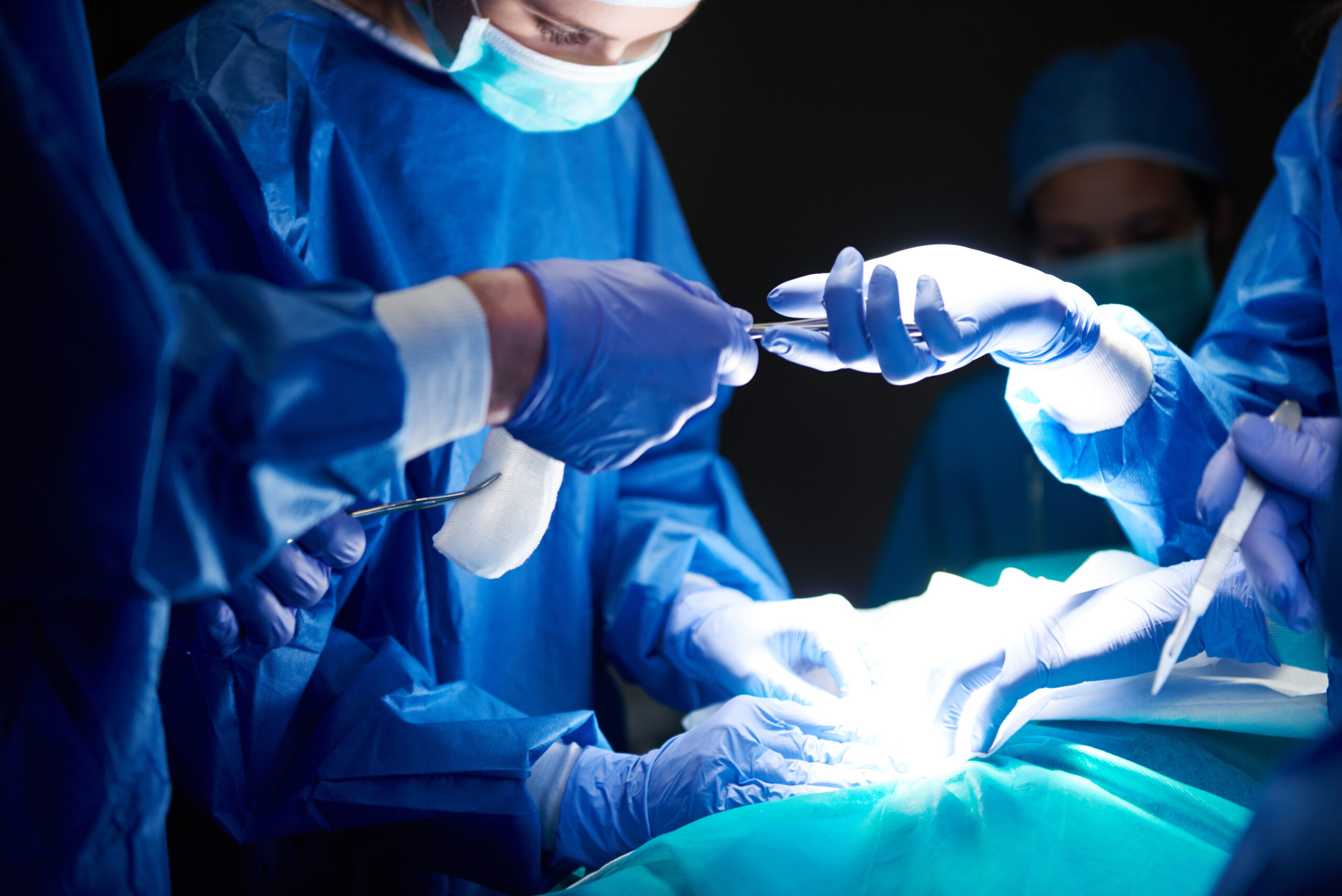
Modern Protocols for Spine Surgery in Varanasi
At Ayaansh Ortho Center, we prioritize patient safety and rapid recovery. Specifically, we design our protocol for Spine Surgery in Varanasi around the Enhanced Recovery After Surgery (ERAS) pathway. This advanced medical approach optimizes every phase of your surgical journey.
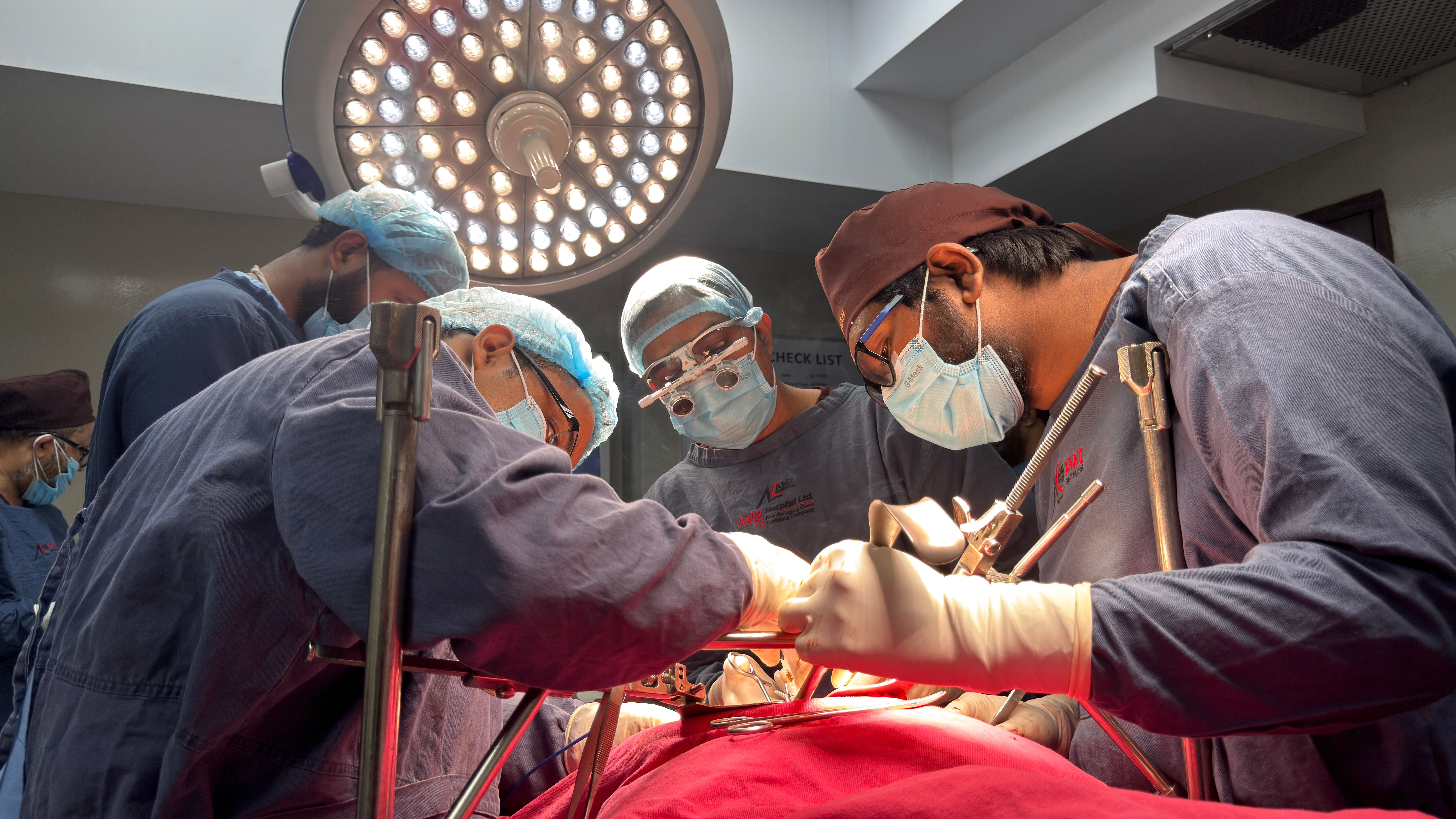
Additionally, we integrate advanced safety technologies to achieve perfect neurological preservation. Whether utilizing computer navigation, high-magnification operating microscopes, or real-time neuromonitoring, our primary goal is to protect your delicate nervous system. This high level of alignment and stabilization precision ensures that physical forces are distributed evenly across your spine. Consequently, this prevents adjacent disc wear, minimizes post-operative pain, and extends the lifespan of your spinal implants.
Part 5: Advanced Rehabilitation and Criteria-Based Physical Therapy
Indeed, your recovery depends heavily on your commitment to physical therapy. During your physical rehabilitation after Spine Surgery in Varanasi, you will follow a customized program designed to restore your spinal mobility and core strength safely.
The Standard Post-Operative Progression Schedule
- Phase 1: Early Mobility & Protection (Weeks 1 to 4): During this first stage, you focus on standing and taking your first steps using a walker, learning to navigate daily tasks safely while observing strict spinal precautions.
- Phase 2: Progressive Strengthening (Weeks 5 to 8): During this intermediate stage, you work toward transitioning to a cane or walking independently, starting gentle core stabilization and stretching.
- Phase 3: Advanced Functional Integration (Weeks 9 to 12): During this final stage, you focus on building advanced core strength, climbing stairs comfortably, and returning to low-impact physical activities.
Phase 1: Homeostasis and Early Mobility (Weeks 1–4)
The primary goals are to manage pain, reduce swelling, activate the deep core muscles, and protect the healing incision.
- Walker-Assisted Walking: The physical therapist helps you stand and take your first steps using a walker on the very first day after surgery.
- Log-Rolling Technique: You must learn to roll your entire body as a single unit when getting in or out of bed to prevent twisting your spine.
- Ankle Pumps: Move your foot up and down rapidly to promote circulation and prevent blood clots.
- Observing Spinal Precautions: You must avoid the “BLT” movements: bending your spine, lifting objects heavier than 5 kilograms, and twisting your back.
Phase 2: Progressive Strengthening (Weeks 5–8)
- Core Stabilization: We work toward strengthening the deep transversus abdominis and multifidus muscles, which act as a natural corset to support your spine.
- Gentle Hamstring Stretching: Tight hamstrings pull on your pelvis, straining the lower back. Gentle stretches help relieve this tension.
- Weaning Off Walker: Transition from a walker to independent walking as your balance and leg strength improve.
Phase 3: Advanced Functional Integration (Weeks 7–12)
- Core and Back Strengthening: Perform bird-dog exercises, pelvic tilts, and glute bridges to rebuild your spinal support system.
- Stair Climbing: Master climbing stairs with confidence, stepping up with the healthy leg first and down with the operated leg.
- Low-Impact Activities: Return to swimming, walking, and stationary cycling.
Frequently Asked Questions (FAQs)
1. Does spine surgery carry a high risk of paralysis?
No. Modern spine surgery is highly safe. Specifically, because we utilize advanced technologies like high-powered microscopes, micro-instruments, and real-time intraoperative neuromonitoring (IONM), the risk of permanent nerve damage or paralysis is exceptionally low (less than 1%).
2. When can I stand and walk after spine surgery?
Under modern Enhanced Recovery After Surgery (ERAS) protocols, most patients stand and walk with the assistance of a physical therapist within 24 hours of their surgery.
3. What is the difference between a spinal decompression and a spinal fusion?
A decompressive surgery (like a microdiscectomy or laminectomy) focuses strictly on removing bone spurs or disc fragments to relieve nerve pressure. In contrast, a spinal fusion involves using screws, rods, and bone grafts to join two or more vertebrae together to stop painful, abnormal movement.
4. How long do I need to stay in the hospital after spine surgery?
For minimally invasive or decompressive procedures (like a microdiscectomy), patients typically stay in the hospital for 1 to 2 days. For complex spinal fusions, a stay of 3 to 4 days is standard before returning home.
5. Why must I avoid smoking before a spinal fusion surgery?
Smoking significantly impairs bone healing. Specifically, nicotine constricts blood vessels and prevents bone-forming cells from fusing the vertebrae together, leading to a high risk of pseudoarthrosis (failure of fusion).
6. What are the “BLT” precautions after spine surgery?
Spinal precautions include avoiding: Bending your spine forward, Lifting objects heavier than 5 kilograms, and Twisting your back. Observing these precautions for the first six weeks protects the healing muscles and bone grafts.
7. When can I return to driving after spine surgery?
You can typically return to driving within 4 to 6 weeks, provided you have regained full control of your limbs, are off all prescription pain medications, and can sit comfortably without pain.
8. What is intraoperative neuromonitoring (IONM)?
IONM is a safety technology that monitors the electrical signals flowing through your spinal cord and nerves during surgery. If a surgical maneuver irritates a nerve, the system alerts the surgeon immediately, allowing him to protect the nerve from injury.
9. Will my spinal flexibility be severely restricted after a fusion?
A single-level lumbar fusion typically causes minimal noticeable loss of flexibility. Specifically, your adjacent joint levels and hips will easily compensate for the lost motion, allowing you to bend and move comfortably.
10. Can I return to sports after spine surgery?
Yes. You can return to low-impact sports such as walking, swimming, cycling, and golf. However, you must avoid high-impact or contact sports like running, football, and weightlifting, especially after a spinal fusion.
11. What causes “Adjacent Level Disease” after spinal fusion?
Fusing a spinal segment transfers all the movement forces to the adjacent discs directly above and below the fusion. Over several years, this increased load can accelerate wear in these adjacent discs, requiring further treatment.
12. What is cauda equina syndrome, and why is it an emergency?
Cauda equina syndrome is a severe neurological emergency caused by massive disc herniation compressing the nerve bundle at the end of the spinal cord. Symptoms include sudden loss of bowel or bladder control, numbness in the saddle area (inner thighs and groin), and progressive leg weakness. It requires immediate surgery within 24 to 48 hours to prevent permanent paralysis.
Expert Care at Ayaansh Ortho Center
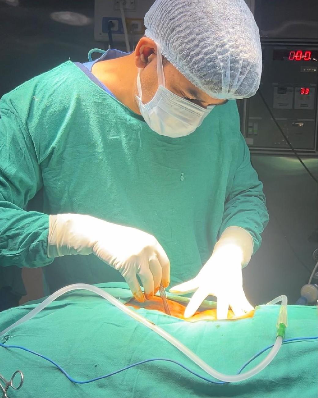
Ayaansh Ortho Center, Opposite Starbucks, Nagwa Lanka, Varanasi, provides comprehensive diagnosis and treatment for shoulder disorders, sports injuries, arthritis, fractures, and spine conditions. Dr. Anand Saurabh (MBBS, MS Orthopaedics), Gold Medalist from IMS BHU with over 10 years of experience, specializes in arthroscopy, arthroplasty, trauma care, and evidence-based orthopedic management. Patients receive personalized treatment plans designed to relieve pain, restore movement, and improve long-term quality of life.
Related Articles
- Back Pain: Causes, Symptoms, Treatment & When to See an Orthopedic Doctor
- Cervical Spondylosis: Causes, Symptoms, Treatment & Prevention
